AChE antibody

AChE antibody detects AChE protein expression by immunohistochemical analysis.
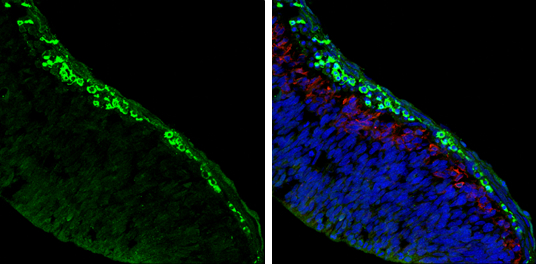
Sample: Frozen sectioned E13.5 Rat brain.
Green: AChE protein stained by AChE antibody (GTX101648) diluted at 1:250.
Red: beta Tubulin 3/ TUJ1, a mature neuron marker, stained by beta Tubulin 3/ TUJ1 antibody [GT11710] (GTX631836) diluted at 1:500.
Blue: Fluoroshield with DAPI (GTX30920).
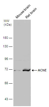
Various tissue extracts (50 μg) were separated by 7.5% SDS-PAGE, and the membrane was blotted with AChE antibody (GTX101648) diluted at 1:500. The HRP-conjugated anti-rabbit IgG antibody (GTX213110-01) was used to detect the primary antibody.
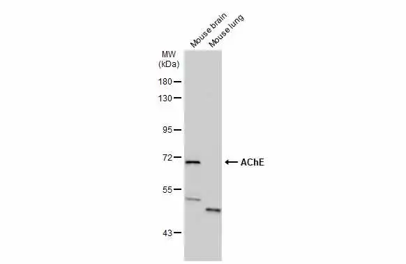
Various tissue extracts (50 μg) were separated by 7.5% SDS-PAGE, and the membrane was blotted with AChE antibody (GTX101648) diluted at 1:500. The HRP-conjugated anti-rabbit IgG antibody (GTX213110-01) was used to detect the primary antibody.
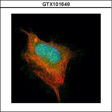
Confocal immunofluorescence analysis (Olympus FV10i) of paraformaldehyde-fixed HeLa, using AChE(GTX101648) antibody (Green) at 1:500 dilution. Alpha-tubulin filaments were labeled with GTX11304 (Red) at 1:2500.
AChE antibody detects AChE protein at nucleus on rat fore brain by immunohistochemical analysis.
Sample: Paraffin-embedded rat fore brain.
AChE antibody (GTX101648) dilution: 1:500.
Antigen Retrieval: Trilogy™ (EDTA based, pH 8.0) buffer, 15min
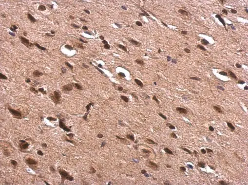
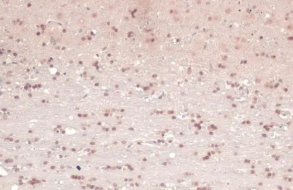
AChE antibody detects AChE protein at cytoplasm and nucleus in rat brain by immunohistochemical analysis.
Sample: Paraffin-embedded rat brain.
AChE antibody (GTX101648) diluted at 1:500.
Antigen Retrieval: Citrate buffer, pH 6.0, 15 min
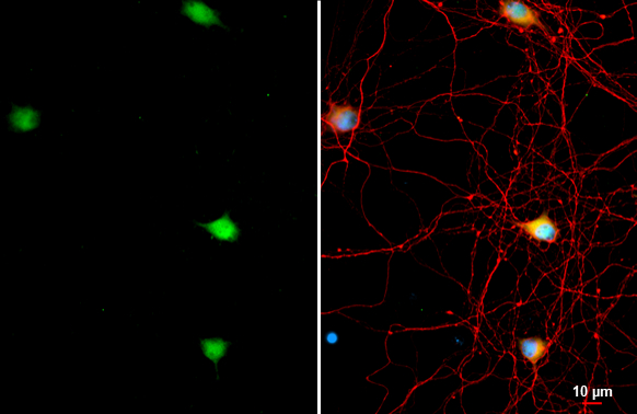
AChE antibody detects AChE protein by immunofluorescent analysis.Sample: DIV9 rat hippocampal neuron and Glia cell cells were fixed in 4% paraformaldehyde at RT for 15 min.Green: AChE stained by AChE antibody (GTX101648) diluted at 1:500.Red: Tau, an axon marker, stained by Tau antibody [GT287] (GTX634809) diluted at 1:500.Blue: Fluoroshield with DAPI (GTX30920).
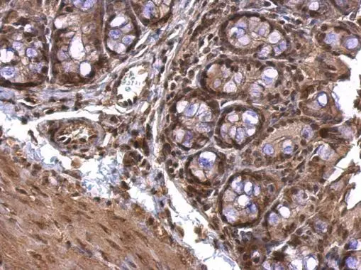
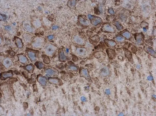
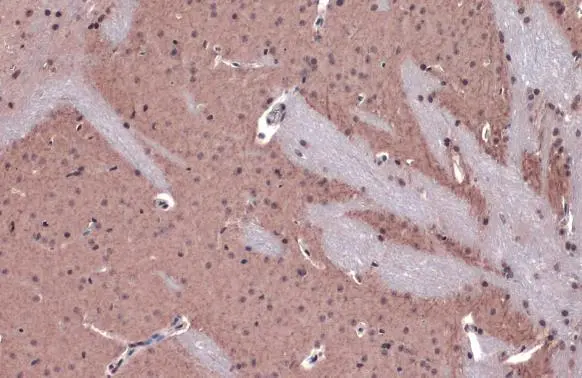
AChE antibody detects AChE protein at nucleus on mouse colon by immunohistochemical analysis.
Sample: Paraffin-embedded mouse colon.
AChE antibody (GTX101648) dilution: 1:500.
Antigen Retrieval: Trilogy™ (EDTA based, pH 8.0) buffer, 15min
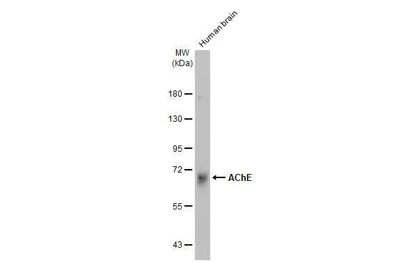
Human tissue extract (30 μg) was separated by 7.5% SDS-PAGE, and the membrane was blotted with AChE antibody (GTX101648) diluted at 1:500. The HRP-conjugated anti-rabbit IgG antibody (GTX213110-01) was used to detect the primary antibody.
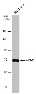
Rat tissue extract (50 μg) was separated by 7.5% SDS-PAGE, and the membrane was blotted with AChE antibody (GTX101648) diluted at 1:500.
AChE antibody detects AChE protein at cell membrane and cytoplasm by immunohistochemical analysis.Sample: Paraffin-embedded mouse brain.AChE stained by AChE antibody (GTX101648) diluted at 1:500.Antigen Retrieval: Citrate buffer, pH 6.0, 15 min
AChE antibody detects AChE protein at nucleus by immunohistochemical analysis.
Sample: Paraffin-embedded mouse brain.
AChE stained by AChE antibody (GTX101648) diluted at 1:500.
Antigen Retrieval: Citrate buffer, pH 6.0, 15 min
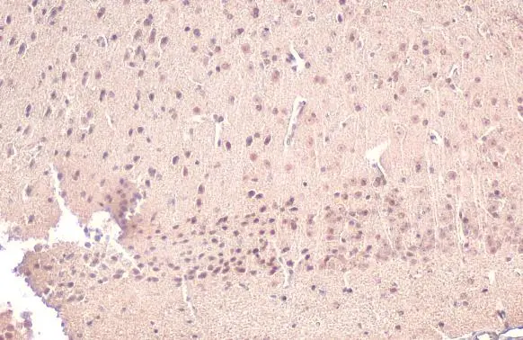
AChE antibody detects AChE protein at nucleus by immunohistochemical analysis.
Sample: Paraffin-embedded rat brain.
AChE stained by AChE antibody (GTX101648) diluted at 1:500.
Antigen Retrieval: Citrate buffer, pH 6.0, 15 min
-
HostRabbit
-
ClonalityPolyclonal
-
IsotypeIgG
-
ApplicationsWB ICC/IF IHC-P IHC-Fr
-
ReactivityHuman, Mouse, Rat