GFP antibody
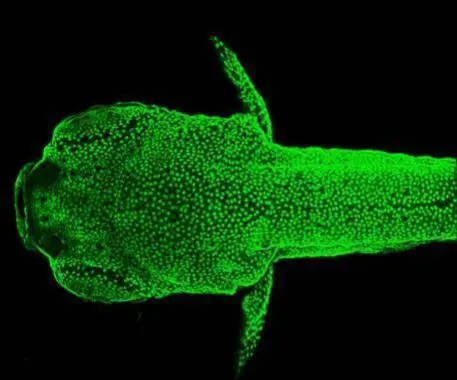
Immunohistochemical analysis (whole mount) of transgenic zebrafish embryo (fixed with ice-cold acetone), using GFP(GTX113617) antibody at 1:200 dilution.
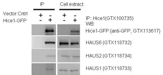
IP-WB assay to show that Hice1 (GTX113617) co-immunoprecipitated with other Augmin components HAUS6 (GTX118732), HAUS2 (GTX118734) and HAUS1 (GTX118733) in U2OS cells. The HRP-conjugated anti-rabbit IgG antibody (GTX213110-01) was used to detect the primary antibody.
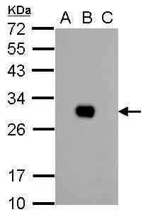
GFP antibody detects GFP protein by western blot analysis.
A. 1 μg 293T whole cell extract
B. 1 μg 293T whole cell extract expressing GFP-tagged protein
C. 1 μg 293T whole cell extract expressing RFP-tagged protein
12% SDS-PAGE
GFP antibody (GTX113617) dilution: 1:10000
The HRP-conjugated anti-rabbit IgG antibody (GTX213110-01) was used to detect the primary antibody.
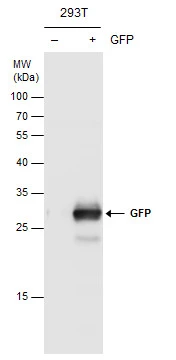
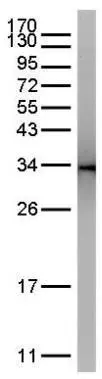
GFP antibody detects GFP protein by Western blot analysis. Non-transfected (-) and GFP-transfected (+) 293T whole cell extracts (30 μg) were separated by 12% SDS-PAGE, and the membrane was blotted with GFP antibody (GTX113617) diluted at 1:10000.
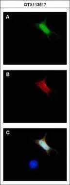
Immunofluorescence analysis of GFP-transfected HeLa. A:GFP is expressed in the tranfected cell. B: The cell expressing GFP can be detected using rabbit anti-GFP ab ( GTX113617, 1:5000) followed by Alexa Fluor 594 (1:500) goat anti-rabbit IgG. C: Merged with DNA probe, the lower cell with no GFP expressed as a negtive control.
GFP was immunoprecipitated from bacterial GFP-expressing lysate using 1 ug of anti-GFP antibody (GTX113617).
The precipitated GFP was detected by GTX113617 diluted at 1:15000.
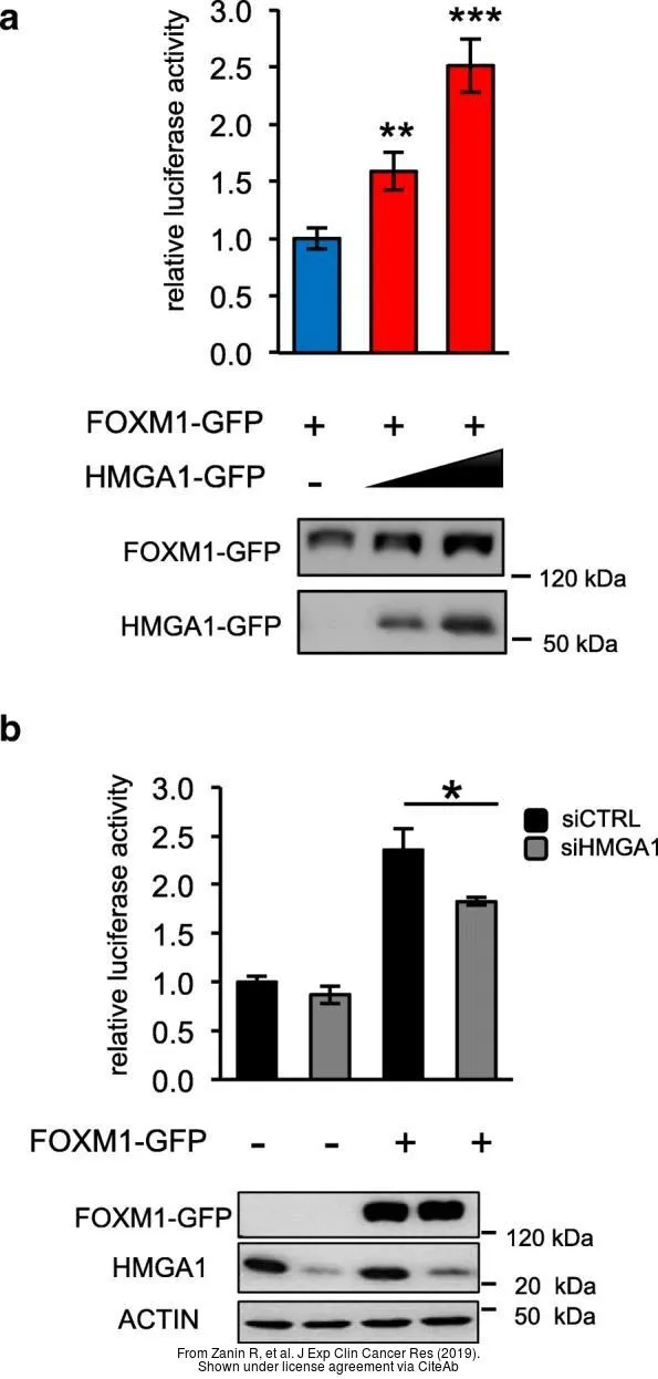
The data was published in the journal J Exp Clin Cancer Res in 2019. PMID: 31311575
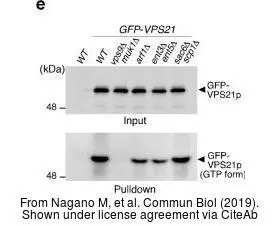
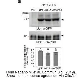
The data was published in the journal Commun Biol in 2019. PMID: 31754649
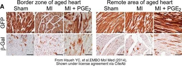
The data was published in the journal EMBO Mol Med in 2014. PMID: 24448489
The data was published in the journal Commun Biol in 2019. PMID: 31754649
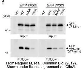
The data was published in the journal Commun Biol in 2019. PMID: 31754649
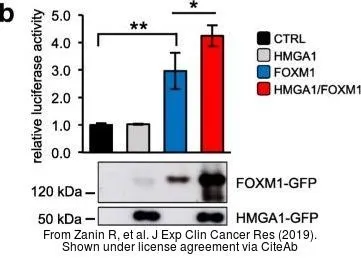
The data was published in the journal J Exp Clin Cancer Res in 2019. PMID: 31311575
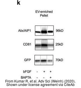
The data was published in the journal Adv Sci (Weinh) in 2020. PMID: 32195080
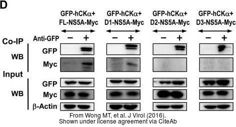
The data was published in the journal J Virol in 2016. PMID: 27489281
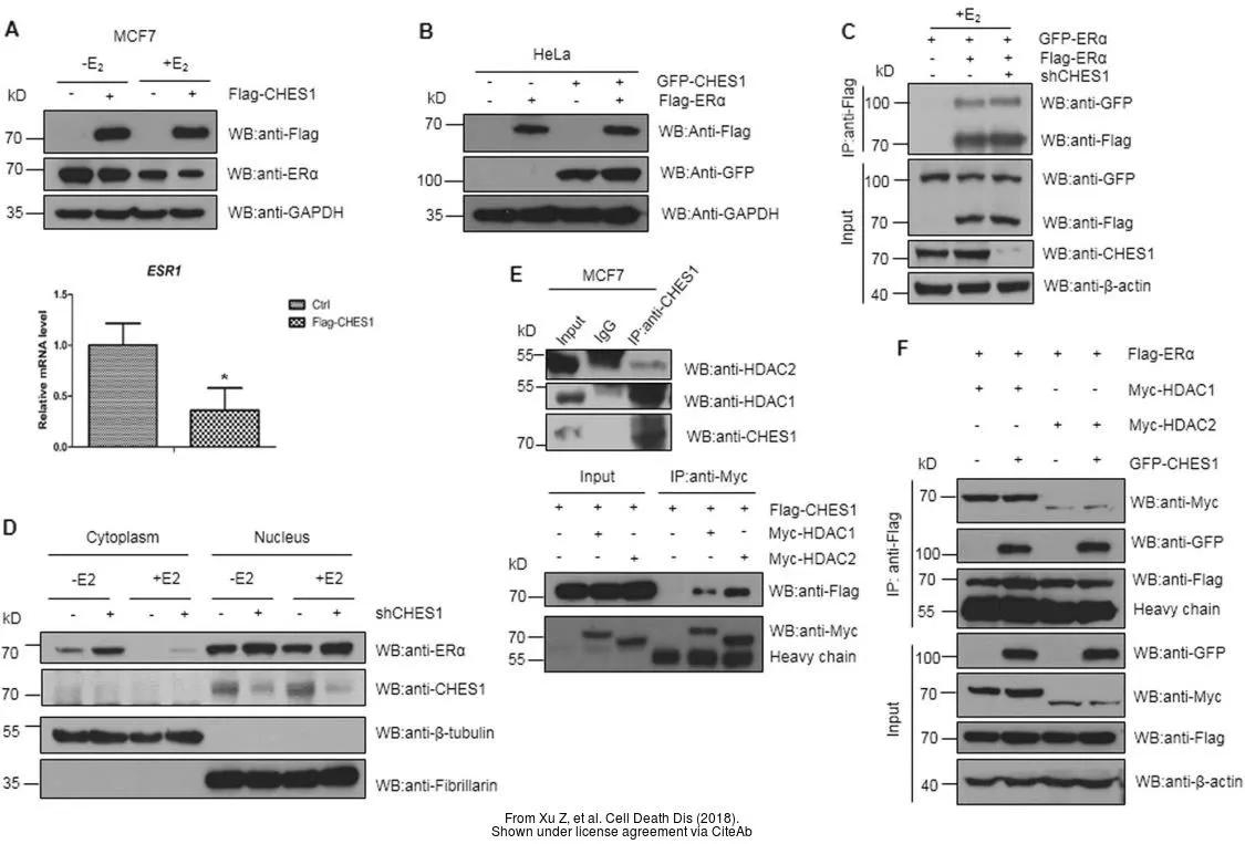
The data was published in the journal Cell Death Dis in 2018.PMID: 29752474
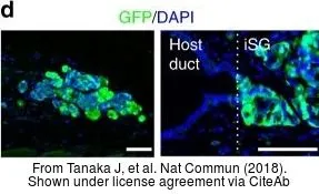
The data was published in the journal Nat Commun in 2018.PMID: 30310071
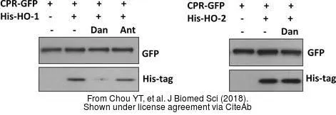
The data was published in the journal J Biomed Sci in 2018.PMID: 29361943
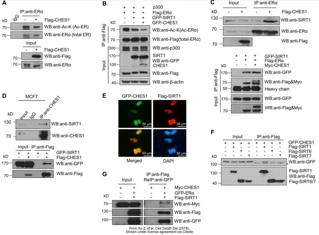
The data was published in the journal Cell Death Dis in 2018.PMID: 29752474
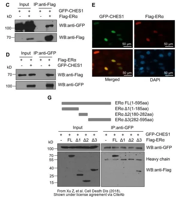
The data was published in the journal Cell Death Dis in 2018.PMID: 29752474
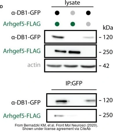
The data was published in the 2020 in Front Mol Neurosci. PMID: 32587503
-
HostRabbit
-
ClonalityPolyclonal
-
IsotypeIgG
-
ApplicationsWB ICC/IF IHC-P IHC-Fr IHC-Wm IP Dot ELISA IHC
-
ReactivitySpecies independent