HDAC2 antibody


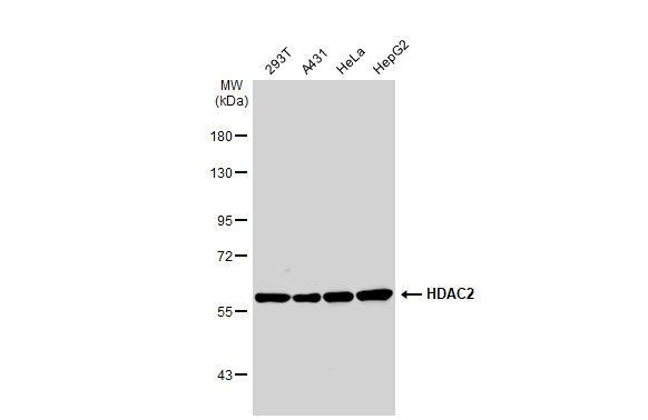
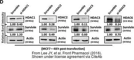
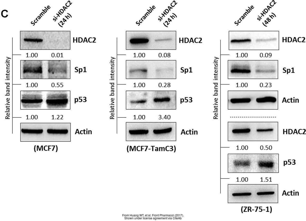
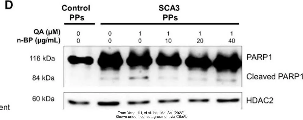
Various whole cell extracts (30 μg) were separated by 7.5% SDS-PAGE, and the membrane was blotted with HDAC2 antibody (GTX109642) diluted at 1:1000. The HRP-conjugated anti-rabbit IgG antibody (GTX213110-01) was used to detect the primary antibody.
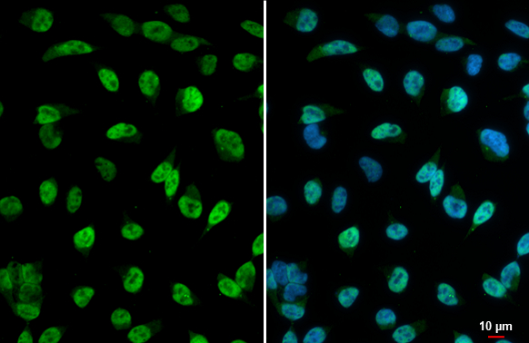
HDAC2 antibody detects HDAC2 protein at nucleus by immunofluorescent analysis.Sample: HeLa cells were fixed in 4% paraformaldehyde at RT for 15 min.Green: HDAC2 stained by HDAC2 antibody (GTX109642) diluted at 1:500.Blue: Fluoroshield with DAPI (GTX30920).
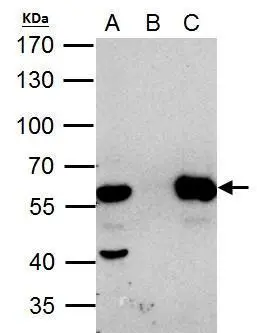
HDAC2 antibody immunoprecipitates HDAC2 protein in IP experiments. IP Sample: HeLa whole cell lysate/extract A. 40 μg HeLa whole cell lysate/extract B. Control with 2 μg of preimmune rabbit IgG C. Immunoprecipitation of HDAC2 protein by 2 μg of HDAC2 antibody (GTX109642) 7.5% SDS-PAGE The immunoprecipitated HDAC2 protein was detected by HDAC2 antibody (GTX109642) diluted at 1:1000. EasyBlot anti-rabbit IgG (GTX221666-01) was used as a secondary reagent.
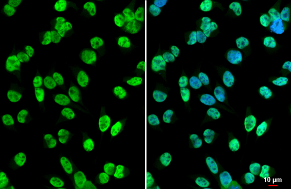
HDAC2 antibody detects HDAC2 protein at nucleus by immunofluorescent analysis.Sample: HeLa cells were fixed in 4% paraformaldehyde at RT for 15 min.Green: HDAC2 stained by HDAC2 antibody (GTX109642) diluted at 1:500.Blue: Fluoroshield with DAPI (GTX30920).
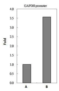
HDAC2 antibody immunoprecipitates HDAC2 protein-DNA complex in ChIP experiments. ChIP Sample: 293T whole cell lysate/extract
A. 5 μg preimmune rabbit IgG
B. 5 μg of HDAC2 antibody (GTX109642)
The precipitated DNA was detected by PCR with primer set targeting to GAPDH promoter.
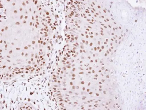
Immunohistochemical analysis of paraffin-embedded Cal27 Xenograft, using HDAC2(GTX109642) antibody at 1:100 dilution.
Antigen Retrieval: Trilogy™ (EDTA based, pH 8.0) buffer, 15min
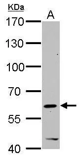
HDAC2 antibody detects HDAC2 protein by western blot analysis.
A. 30 μg C2C12 whole cell lysate/extract
7.5% SDS-PAGE
HDAC2 antibody (GTX109642) dilution: 1:1000
The HRP-conjugated anti-rabbit IgG antibody (GTX213110-01) was used to detect the primary antibody.
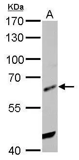
HDAC2 antibody detects HDAC2 protein by western blot analysis.
A. 30 μg Rat2 whole cell lysate/extract
7.5% SDS-PAGE
HDAC2 antibody (GTX109642) dilution: 1:1000
The HRP-conjugated anti-rabbit IgG antibody (GTX213110-01) was used to detect the primary antibody.
-
HostRabbit
-
ClonalityPolyclonal
-
IsotypeIgG
-
ApplicationsWB ICC/IF IHC-P IP ChIP assay
-
ReactivityHuman, Mouse, Rat, Xenopus tropicalis