HIF1 alpha antibody


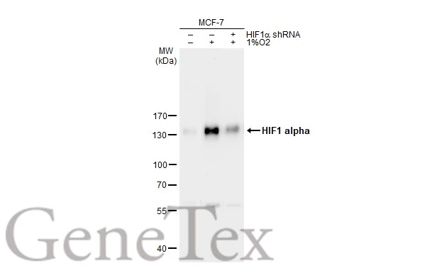
Untreated (–) and treated (+) MCF-7 whole cell extracts (30 μg) were separated by 7.5% SDS-PAGE, and the membrane was blotted with HIF1 alpha antibody (GTX127309) diluted at 1:1000. The HRP-conjugated anti-rabbit IgG antibody (GTX213110-01) was used to detect the primary antibody.
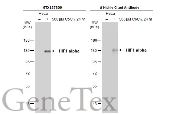
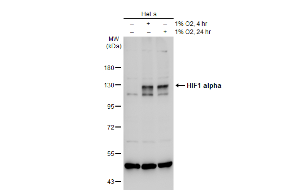
Untreated (–) and treated (+) HeLa whole cell extracts (30 μg) were separated by 7.5% SDS-PAGE, and the membrane was blotted with HIF1 alpha antibody (GTX127309) diluted at 1:1000. The HRP-conjugated anti-rabbit IgG antibody (GTX213110-01) was used to detect the primary antibody.
*The competitor is not affiliated with GeneTex and does not endorse this product.
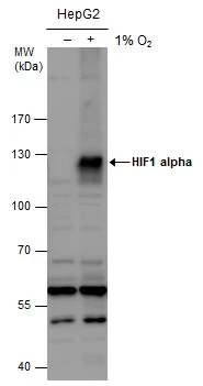
HIF1 alpha antibody detects HIF1 alpha protein by western blot analysis. Un-treated (-) and treated (+, 1% O2 treatment for 24hr) HepG2 whole cell extracts (30 μg) were separated by 7.5% SDS-PAGE, and the membrane was blotted with HIF1 alpha antibody (GTX127309) diluted at 1:1000. The HRP-conjugated anti-rabbit IgG antibody (GTX213110-01) was used to detect the primary antibody.
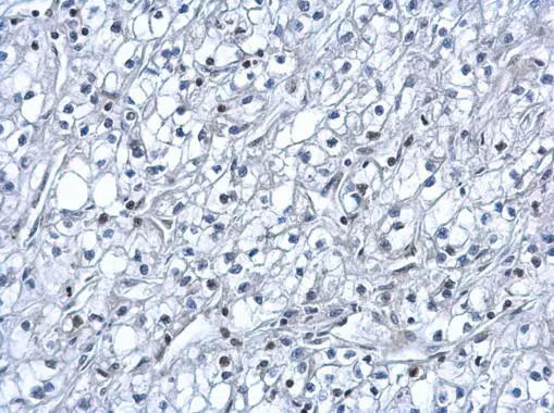
HIF1 alpha antibody detects HIF1 alpha protein at nucleus on human kidney cancer by immunohistochemical analysis.
Sample: Paraffin-embedded human kidney cancer.
HIF1 alpha antibody (GTX127309) dilution: 1:500.
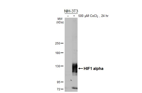
Untreated (–) and treated (+) NIH-3T3 whole cell extracts (30 μg) were separated by 5% SDS-PAGE, and the membrane was blotted with HIF1 alpha antibody (GTX127309) diluted at 1:1000. The HRP-conjugated anti-rabbit IgG antibody (GTX213110-01) was used to detect the primary antibody.

Untreated (–) and treated (+) Rat2 whole cell extracts (30 μg) were separated by 5% SDS-PAGE, and the membrane was blotted with HIF1 alpha antibody (GTX127309) diluted at 1:1000. The HRP-conjugated anti-rabbit IgG antibody (GTX213110-01) was used to detect the primary antibody.

Untreated (–) and treated (+) HeLa whole cell extracts (30 μg) were separated by 5% SDS-PAGE, and the membrane was blotted with HIF1 alpha antibody (GTX127309) diluted at 1:1000. The HRP-conjugated anti-rabbit IgG antibody (GTX213110-01) was used to detect the primary antibody.
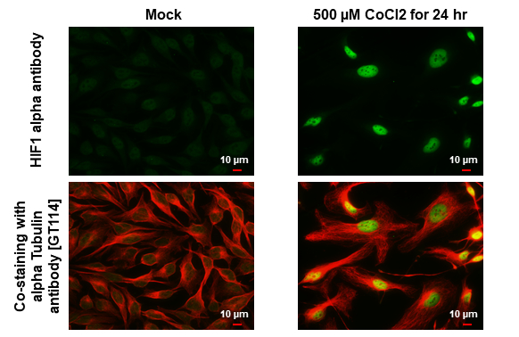
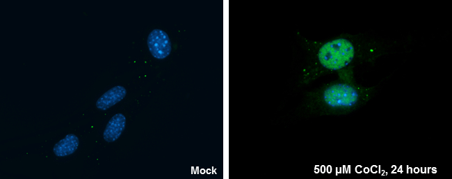
HIF1 alpha antibody detects HIF1 alpha protein by immunofluorescent analysis.Sample: Mock and treated HeLa cells were fixed in 4% paraformaldehyde at RT for 15 min.Green: HIF1 alpha stained by HIF1 alpha antibody (GTX127309) diluted at 1:500.Red: alpha Tubulin, a cytoskeleton marker, stained by alpha Tubulin antibody [GT114] (GTX628802) diluted at 1:1000.
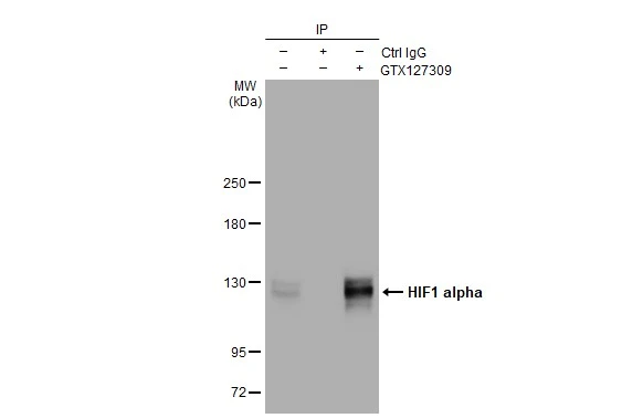
Immunoprecipitation of HIF1 alpha protein from HeLa whole cell extracts treated with 500 μM CoCl2 for 24 hr using 5 μg of HIF1 alpha antibody (GTX127309).
Western blot analysis was performed using HIF1 alpha antibody (GTX127309).
EasyBlot HRP-conjugated anti rabbit IgG antibody (GTX221666-01) was used to detect the primary antibody.
Untreated (–) and treated (+) HeLa whole cell extracts (30 μg) were separated by 7.5% SDS-PAGE, and the membrane was blotted with HIF1 alpha antibody (GTX127309) diluted at 1:1000. The HRP-conjugated anti-rabbit IgG antibody (GTX213110-01) was used to detect the primary antibody.
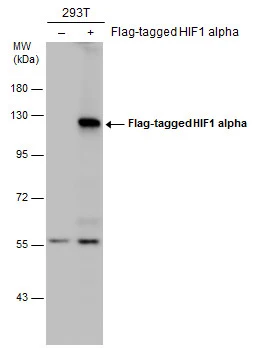
Non-transfected (–) and transfected (+) 293T whole cell extracts (30 μg) were separated by 7.5% SDS-PAGE, and the membrane was blotted with HIF1 alpha antibody (GTX127309) diluted at 1:5000. The HRP-conjugated anti-rabbit IgG antibody (GTX213110-01) was used to detect the primary antibody.
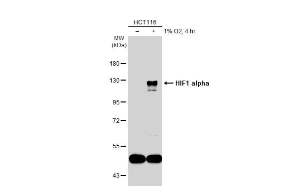
Untreated (–) and treated (+) HCT116 whole cell extracts (30 μg) were separated by 7.5% SDS-PAGE, and the membrane was blotted with HIF1 alpha antibody (GTX127309) diluted at 1:1000. The HRP-conjugated anti-rabbit IgG antibody (GTX213110-01) was used to detect the primary antibody.
HIF1 alpha antibody detects HIF1 alpha protein at nucleus by immunofluorescent analysis.
Sample: NIH/3T3 cells were fixed in 4% paraformaldehyde at RT for 15 min.
Green: HIF1 alpha protein stained by HIF1 alpha antibody (GTX127309) diluted at 1:200.
Blue: Hoechst 33342 staining.
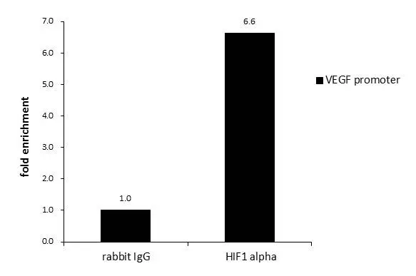
ChIP was performed with HepG2 chromatin extract and 5 μg of either normal rabbit IgG or anti-HIF1 alpha antibody. The precipitated DNA was detected by PCR with primer set targeting to VEGF promoter.
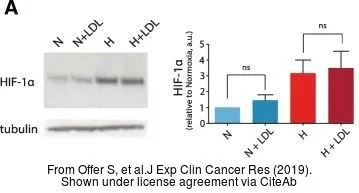
The data was published in the journal J Exp Clin Cancer Res in 2019. PMID: 31174567
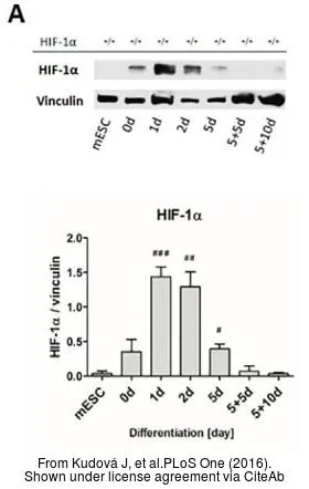
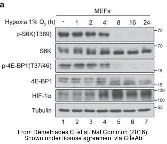
The data was published in the journal PLoS One in 2016. PMID: 27355368
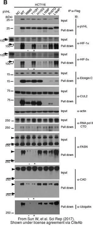
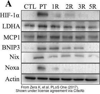
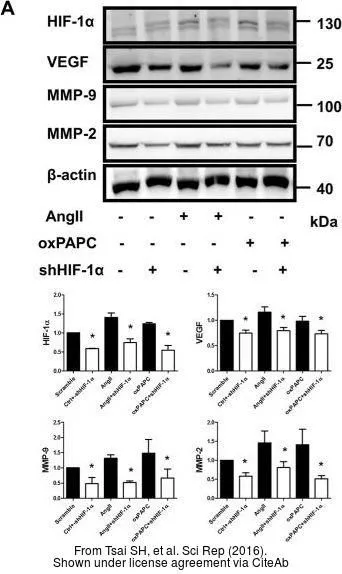
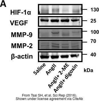
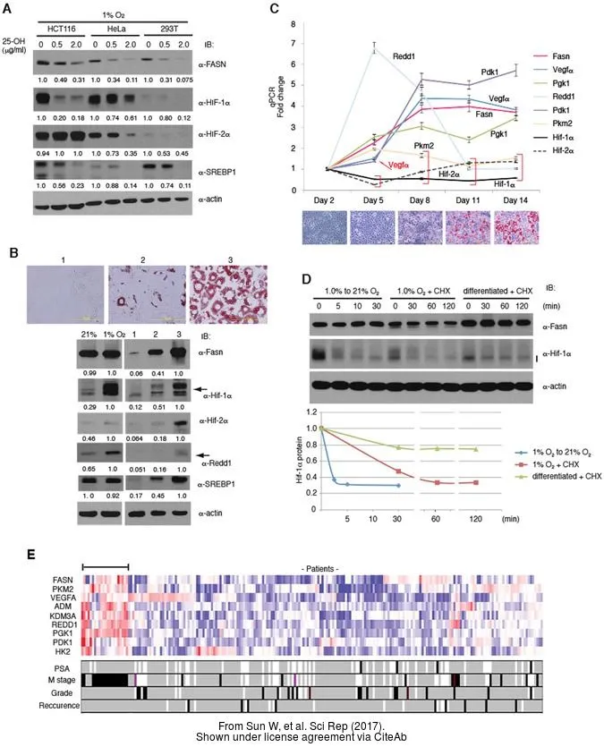
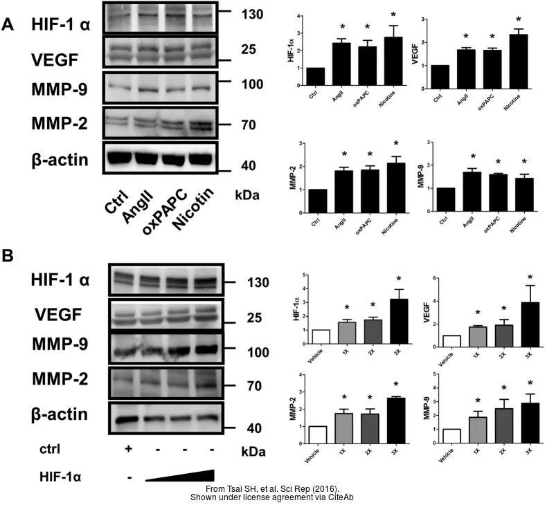
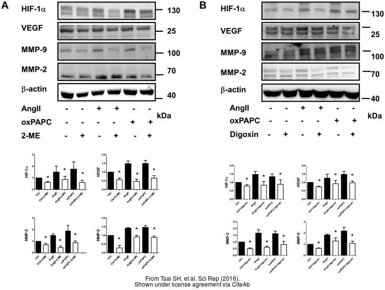
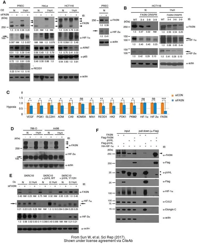
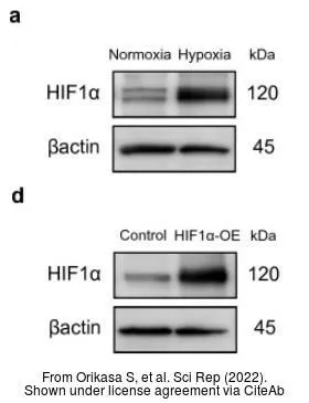
The data was published in the journal Sci Rep in 2017. PMID: 28775317
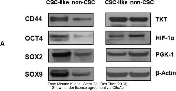
The data was published in the journal Stem Cell Res Ther in 2013. PMID: 24423398
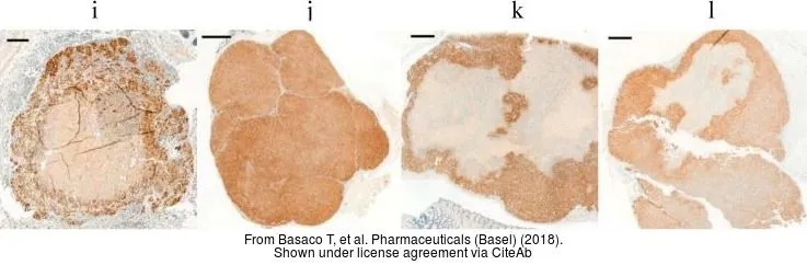
The data was published in the journal Pharmaceuticals (Basel) in 2018.PMID: 30487460
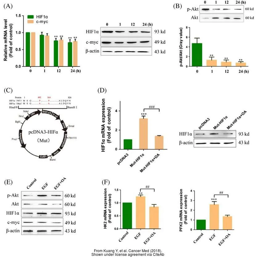
The data was published in the journal Cancer Med in 2018.PMID: 29533007
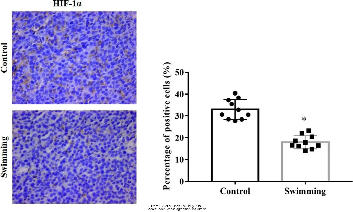
The data was published in the 2022 in Open Life Sci. PMID: 35291563
-
HostRabbit
-
ClonalityPolyclonal
-
IsotypeIgG
-
ApplicationsWB ICC/IF IHC-P IHC-Fr IP ChIP assay
-
ReactivityHuman, Mouse, Rat, Rabbit, Bovine