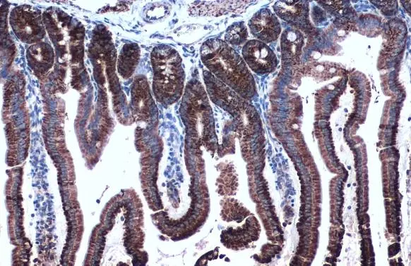
HSP60 antibody

HSP60 antibody detects HSP60 protein at mitochondrion on mouse colon by immunohistochemical analysis.
Sample: Paraffin-embedded mouse colon.
HSP60 antibody (GTX110089) dilution: 1:500.
Antigen Retrieval: Trilogy™ (EDTA based, pH 8.0) buffer, 15min
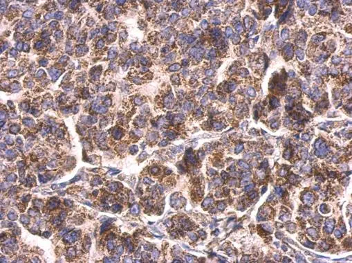
HSP60 antibody detects HSP60 protein at mitochondria on human breast carcinoma by immunohistochemical analysis.
Sample: Paraffin-embedded human breast carcinoma.
HSP60 antibody (GTX110089) dilution: 1:500.
Antigen Retrieval: Trilogy™ (EDTA based, pH 8.0) buffer, 15min
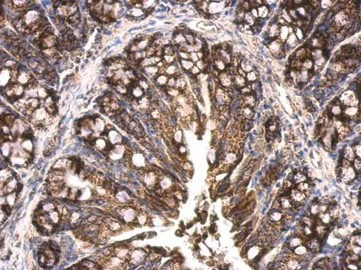
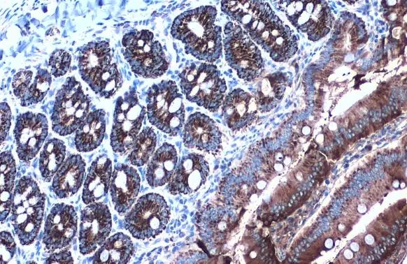
HSP60 antibody detects HSP60 protein at mitochondria on human colon carcinoma by immunohistochemical analysis.
Sample: Paraffin-embedded human colon carcinoma.
HSP60 antibody (GTX110089) dilution: 1:500.
Antigen Retrieval: Trilogy™ (EDTA based, pH 8.0) buffer, 15min
HSP60 antibody detects HSP60 protein at mitochondria by immunohistochemical analysis.Sample: Paraffin-embedded mouse intestine.HSP60 stained by HSP60 antibody (GTX110089) diluted at 1:1000.Antigen Retrieval: Citrate buffer, pH 6.0, 15 min
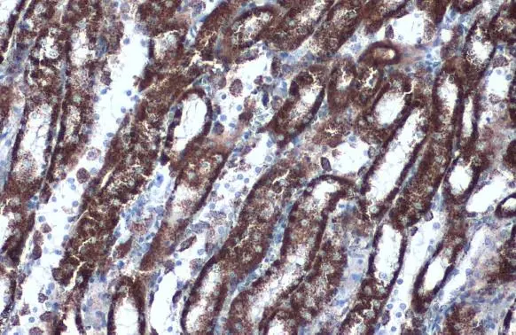
HSP60 antibody detects HSP60 protein at mitochondria by immunohistochemical analysis.Sample: Paraffin-embedded rat duodenum.HSP60 stained by HSP60 antibody (GTX110089) diluted at 1:500.Antigen Retrieval: Citrate buffer, pH 6.0, 15 min
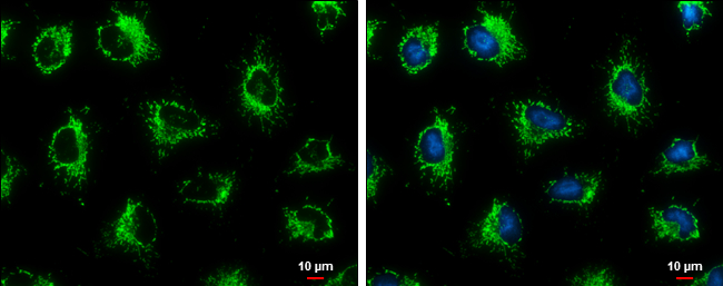
HSP60 antibody detects HSP60 protein at mitochondria by immunofluorescent analysis.
Sample: HeLa cells were fixed in ice-cold MeOH for 5 min.
Green: HSP60 protein stained by HSP60 antibody (GTX110089) diluted at 1:500.
Blue: Hoechst 33342 staining.
Scale bar = 10 μm.
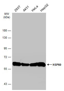
Various whole cell extracts (30 μg) were separated by 7.5% SDS-PAGE, and the membrane was blotted with HSP60 antibody (GTX110089) diluted at 1:10000. The HRP-conjugated anti-rabbit IgG antibody (GTX213110-01) was used to detect the primary antibody.
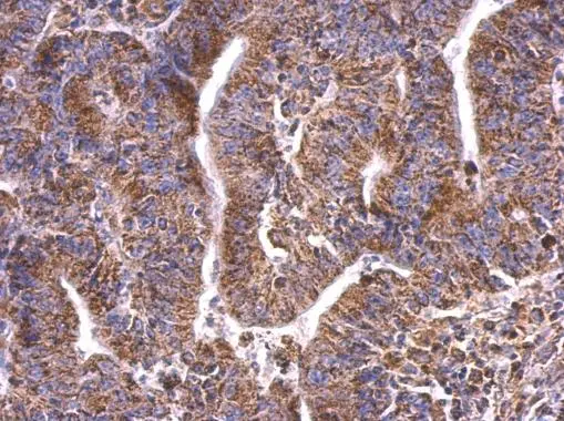
HSP60 antibody detects HSP60 protein at mitochondria by immunohistochemical analysis.Sample: Paraffin-embedded rat kidney.HSP60 stained by HSP60 antibody (GTX110089) diluted at 1:1000.Antigen Retrieval: Citrate buffer, pH 6.0, 15 min
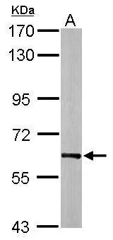
Sample (20 μg of whole cell lysate)
A: mouse brain
7.5% SDS PAGE
GTX110089 diluted at 1:20000
The HRP-conjugated anti-rabbit IgG antibody (GTX213110-01) was used to detect the primary antibody.
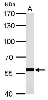
HSP60 antibody detects HSPD1 protein by western blot analysis.
A. 50 μg rat brain lysate/extract
7.5% SDS-PAGE
HSP60 antibody (GTX110089) dilution: 1:10000
The HRP-conjugated anti-rabbit IgG antibody (GTX213110-01) was used to detect the primary antibody.
Drosophila Hsp60A is the homolog of mammalian Hsp60. (Gene ID: 32045/ UniProt: O02649)
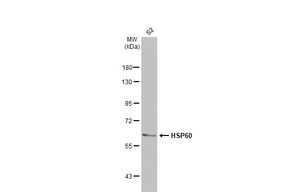
Whole cell extract (30 μg) was separated by 7.5% SDS-PAGE, and the membrane was blotted with HSP60 antibody (GTX110089) diluted at 1:1000. The HRP-conjugated anti-rabbit IgG antibody (GTX213110-01) was used to detect the primary antibody.
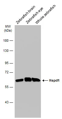
Various tissue extracts (30 μg) were separated by 7.5% SDS-PAGE, and the membrane was blotted with HSP60 antibody (GTX110089) diluted at 1:1000. The HRP-conjugated anti-rabbit IgG antibody (GTX213110-01) was used to detect the primary antibody.
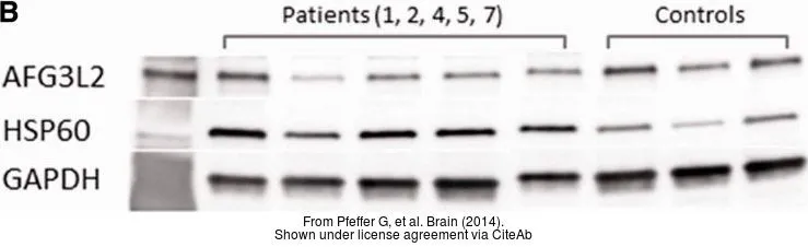
The data was published in the journal Brain in 2014. PMID: 24727571
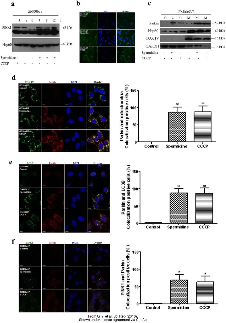
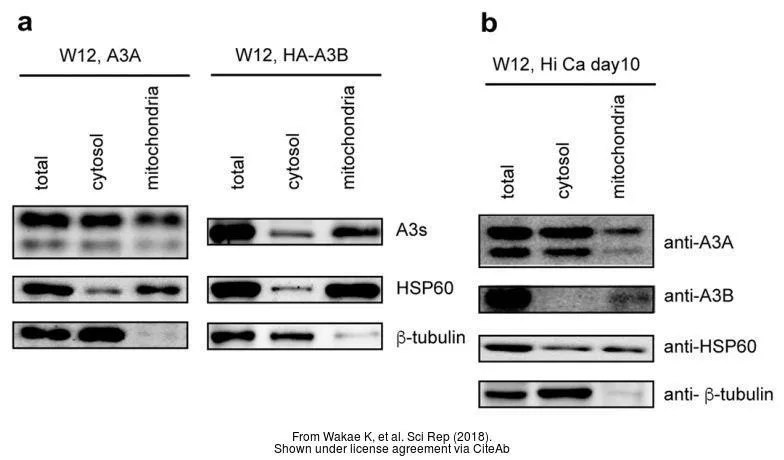
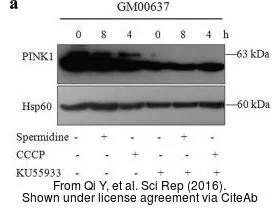
The data was published in the journal Sci Rep in 2016. PMID: 27089984
The data was published in the journal Sci Rep in 2016. PMID: 27089984
-
HostRabbit
-
ClonalityPolyclonal
-
IsotypeIgG
-
ApplicationsWB ICC/IF IHC-P ELISA
-
ReactivityHuman, Mouse, Rat, Zebrafish, Drosophila, Hamster