GeneTex
  • Country / Location Selection

United States (US)
ATR antibody

Anti-ATR antibody used in Western Blot (WB). GTX128146
Anti-ATR antibody used in Immunocytochemistry/ Immunofluorescence (ICC/IF). GTX128146
Anti-ATR antibody used in IHC (Paraffin sections) (IHC-P). GTX128146
Anti-ATR antibody used in Western Blot (WB). GTX128146
Anti-ATR antibody used in IHC (Paraffin sections) (IHC-P). GTX128146
Anti-ATR antibody used in Western Blot (WB). GTX128146
Anti-ATR antibody used in Immunoprecipitation (IP). GTX128146
Anti-ATR antibody used in Western Blot (WB). GTX128146

Cat. No. GTX128146

Host

Rabbit

Clonality

Polyclonal

Isotype

IgG

Application

WB, ICC/IF, IHC-P, IP

Reactivity

Human, Mouse
Package
100 μl ($399),
25 μl ($169)
PRODUCT

Summary

ATR antibody detects the serine/threonine-protein kinase ATR (Ataxia Telangiectasia and Rad3-related protein) (also known as FRAP-related protein 1 (FRP1)). ATR (301 kDa) is a key component of the DNA damage response machinery and is activated by ssDNA, leading to cell cycle arrest while DNA damage is repaired. It also plays a critical role in stabilizing the genome during DNA replication by preventing premature onset of mitosis. This ATR antibody is a cited rabbit polyclonal validated for multiple applications.

APPLICATION

Application Note

*Optimal dilutions/concentrations should be determined by the researcher.
Application Recommended Dilution
WB 1:500-1:3000
ICC/IF 1:100-1:5000
IHC-P 1:100-1:1000
IP 1:100-1:500
Not tested in other applications.

Calculated MW

301 kDa. ( Note )

Positive Control

293T , A431 , HeLa , HepG2 , Raji nuclear extract

PROPERTIES

Form

Liquid

Buffer

PBS, 1% BSA, 20% Glycerol

Preservative

0.025% ProClin 300

Storage

Store as concentrated solution. Centrifuge briefly prior to opening vial. For short-term storage (1-2 weeks), store at 4ºC. For long-term storage, aliquot and store at -20ºC or below. Avoid multiple freeze-thaw cycles.

Concentration

1 mg/ml (Please refer to the vial label for the specific concentration.)

Antigen Species

Human

Immunogen

Carrier-protein conjugated synthetic peptide encompassing a sequence within the C-terminus region of human ATR. The exact sequence is proprietary.

Purification

Purified by antigen-affinity chromatography.

Conjugation

Unconjugated

RRID

AB_2885716

Note

For laboratory research use only. Not for any clinical, therapeutic, or diagnostic use in humans or animals. Not for animal or human consumption.

Purchasers shall not, and agree not to enable third parties to, analyze, copy, reverse engineer or otherwise attempt to determine the structure or sequence of the product.

TARGET

Synonyms

ATR serine/threonine kinase , FCTCS , FRP1 , MEC1 , SCKL , SCKL1

Cellular Localization

Nucleus

Background

The protein encoded by this gene belongs the PI3/PI4-kinase family, and is most closely related to ATM, a protein kinase encoded by the gene mutated in ataxia telangiectasia. This protein and ATM share similarity with Schizosaccharomyces pombe rad3, a cell cycle checkpoint gene required for cell cycle arrest and DNA damage repair in response to DNA damage. This kinase has been shown to phosphorylate checkpoint kinase CHK1, checkpoint proteins RAD17, and RAD9, as well as tumor suppressor protein BRCA1. Mutations of this gene are associated with Seckel syndrome. An alternatively spliced transcript variant of this gene has been reported, however, its full length nature is not known. Transcript variants utilizing alternative polyA sites exist. [provided by RefSeq]

Database

Research Area

DATA IMAGES

Anti-ATR antibody used in Western Blot (WB). GTX128146

GTX128146 WB Image

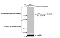
Untreated (–) and treated (+) HeLa whole cell extracts (30 μg) were separated by 5% SDS-PAGE, and the membrane was blotted with ATR antibody (GTX128146) diluted at 1:500. The HRP-conjugated anti-rabbit IgG antibody (GTX213110-01) was used to detect the primary antibody, and the signal was developed with Trident ECL plus-Enhanced.

Anti-ATR antibody used in Western Blot (WB). GTX128146

GTX128146 WB Image

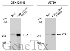
Raji whole cell and nuclear extracts (30 μg) were separated by 5% SDS-PAGE, and the membranes were blotted with ATR antibody (GTX128146) diluted at 1:500 and competitor's antibody (CST#2790) diluted at 1:1000. The HRP-conjugated anti-rabbit IgG antibody (GTX213110-01) was used to detect the primary antibody.
*The competitor is not affiliated with GeneTex and does not endorse this product.

Anti-ATR antibody used in Immunocytochemistry/ Immunofluorescence (ICC/IF). GTX128146

GTX128146 ICC/IF Image

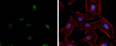
ATR antibody detects ATR protein at nucleolus by immunofluorescent analysis.
Sample: HeLa cells were fixed in 4% paraformaldehyde at RT for 15 min.
Green: ATR protein stained by ATR antibody (GTX128146) diluted at 1:5000.
Red: phalloidin, a cytoskeleton marker, diluted at 1:200.
Blue: Hoechst 33342 staining.
Scale bar = 10 μm.

Anti-ATR antibody used in IHC (Paraffin sections) (IHC-P). GTX128146

GTX128146 IHC-P Image

ATR antibody detects ATR protein at nucleus on mouse testis by immunohistochemical analysis.
Sample: Paraffin-embedded mouse testis.
ATR antibody (GTX128146) dilution: 1:500.

Antigen Retrieval: Trilogy™ (EDTA based, pH 8.0) buffer, 15min

Anti-ATR antibody used in Western Blot (WB). GTX128146

GTX128146 WB Image

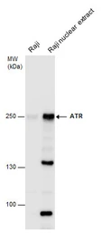
ATR antibody detects ATR protein by western blot analysis. Raji whole cell extracts and nuclear extracts (30 μg) were separated by 5% SDS-PAGE, and the membrane was blotted with ATR antibody (GTX128146) at a dilution of 1:500. The HRP-conjugated anti-rabbit IgG antibody (GTX213110-01) was used to detect the primary antibody.

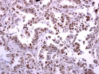
Anti-ATR antibody used in IHC (Paraffin sections) (IHC-P). GTX128146

GTX128146 IHC-P Image

ATR antibody detects ATR protein at nucleus on human lung carcinoma by immunohistochemical analysis.
Sample: Paraffin-embedded human lung carcinoma.
ATR antibody (GTX128146) diluted at 1:250.

Antigen Retrieval: Trilogy™ (EDTA based, pH 8.0) buffer, 15min

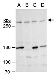
Anti-ATR antibody used in Western Blot (WB). GTX128146

GTX128146 WB Image

ATR antibody detects ATR protein by Western blot analysis.
A. 30 μg 293T whole cell extract
B. 30 μg A431 whole cell extract
C. 30 μg HeLa whole cell extract
D. 30 μg HepG2 whole cell extract
5 % SDS-PAGE
ATR antibody (GTX128146) dilution: 1:500

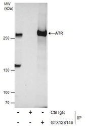
Anti-ATR antibody used in Immunoprecipitation (IP). GTX128146

GTX128146 IP Image

Immunoprecipitation of ATR protein from HCT-116 whole cell extracts treated with Hydroxyurea for 6 hr using 5 μg of ATR antibody (GTX128146).
Western blot analysis was performed using ATR antibody (GTX128146).
EasyBlot anti-Rabbit IgG (GTX221666-01) was used as a secondary reagent.

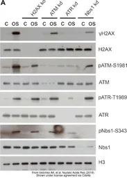
Anti-ATR antibody used in Western Blot (WB). GTX128146

GTX128146 WB Image

The data was published in the journal Nucleic Acids Res in 2019. PMID: 31114877

REFERENCE

REVIEW

There are currently no reviews for ATR antibody (GTX128146). Be the first to share your experience with this product.
SDS
PBS.pdf
Glycerol.pdf
Thimerosal.pdf
Package List Price ($)
$ 399
$ 169