AKT antibody

WB analysis of PDGF treated and untreated NIH-3T3 whole cell lysate using GTX28805 AKT antibody.
Green : Primary antibody
Red : Akt (phospho Ser473)
Lane 1 : Untreated whole cell lysate
Lane 2 : PDGF-treated whole cell lysate
Loading : 35 μg
Dilution : 1:1000

ICC/IF analysis of PFA-fixed rat neonatal cardiomyocytes using GTX28805 AKT antibody.
Green : Primary antibody
Red : Actin
Dilution : 1:80
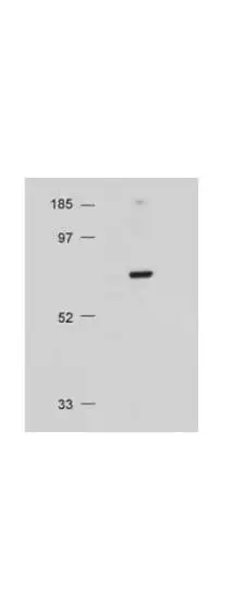
WB analysis of NIH-3T3 whole cell lysate using GTX28805 AKT antibody.
Loading : 20 μg
Dilution : 1:500
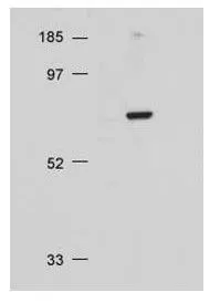
Western blot using Akt antibody at 1:500 for overnight at 4ºC on 20 ug NIH/3T3 whole cell lysate.
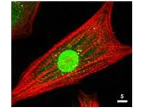
ICC/IF of the Akt antibody is used at 1: 80 for 1hr at RT on cultured neonatal rat cardiomyocytes (Green). The red is Texas-red phalloidin that labels actin filaments.

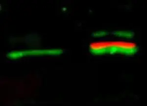
Western Blot of simultaneous detection of unphosphorylated and phosphorylated Rabbit Anti-AKT antibody (GTX28805). Lane 1: unstimulated NIH/3T3 lysates contain inactive unphosphorylated Akt1, green band. Lane 2: PDGF stimulated NIH/3T3 lysate contains both inactive (green band) and activated phosphorylated Akt1 (red band). Load: 35 μg per lane. Primary antibody: rabbit anti-Akt (pan) and mouse anti-Akt pS473 specific antibodies at 1:1000 for overnight at 4ºC. Secondary antibody: Flourescent 549 conjugated anti-rabbit IgG (green) and flourescent 649 conjugated anti-mouse IgG (red) secondary antibodies at 1:10,000 for 45 min at RT.
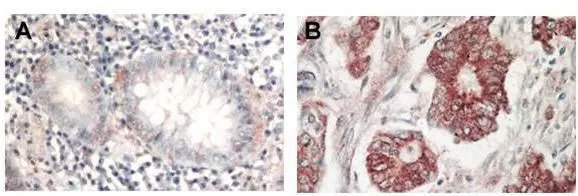
Immunohistochemical analysis of formalin-fixed paraffin-embedded tissue, A: Normal colon tissue, B: Colon tumor tissue, using AKT(GTX28805) antibody at 1:1,000 dilution for 1 h at RT. Peroxidase rabbit secondary antibody at 1:10,000 for 45 min at RT. Localization: AKT is nuclear. Staining: AKT as precipitated red signal with hematoxylin purple nuclear counterstain.
-
HostRabbit
-
ClonalityPolyclonal
-
IsotypeIgG
-
ApplicationsWB ICC/IF IHC-P FCM ELISA
-
ReactivityHuman, Mouse, Rat, Chicken