APPLICATION
Application Note
*Optimal dilutions/concentrations should be determined by the researcher.
| Application |
Recommended Dilution |
| 1:500-1:2,000 |
| 2 μg/mL |
| 1:100 |
| Assay dependent |
| Assay dependent |
Not tested in other applications.
Calculated MW
PROPERTIES
Form
Liquid
Buffer
PBS, 1 mg/ml BSA
Preservative
0.05% sodium azide
Storage
Store as concentrated solution. Centrifuge briefly prior to opening vial. For short-term storage (1-2 weeks), store at 4ºC. For long-term storage, aliquot and store at -20ºC or below. Avoid multiple freeze-thaw cycles.
Concentration
2 mg/ml (Please refer to the vial label for the specific concentration.)
Antigen Species
Human
Immunogen
Synthetic peptide: RGERTAFIKDQSAL, corresponding to amino acids 780-793 of Human Furin Convertase.
Purification
Purified by antigen-affinity chromatography
Conjugation
Unconjugated
Note
For laboratory research use only. Not for any clinical, therapeutic, or diagnostic use in humans or animals. Not for animal or human consumption.
Purchasers shall not, and agree not to enable third parties to, analyze, copy, reverse engineer or otherwise attempt to determine the structure or sequence of the product.
TARGET
Synonyms
FUR,FURIN,PACE,PCSK3,SPC1,furin, paired basic amino acid cleaving enzyme,Furin
Cellular Localization
Golgi apparatus,Trans-Golgi network membrane,Cell membrane,Secreted,Endosome membrane
Background
This gene encodes a member of the subtilisin-like proprotein convertase family, which includes proteases that process protein and peptide precursors trafficking through regulated or constitutive branches of the secretory pathway. It encodes a type 1 membrane bound protease that is expressed in many tissues, including neuroendocrine, liver, gut, and brain. The encoded protein undergoes an initial autocatalytic processing event in the ER and then sorts to the trans-Golgi network through endosomes where a second autocatalytic event takes place and the catalytic activity is acquired. The product of this gene is one of the seven basic amino acid-specific members which cleave their substrates at single or paired basic residues. Some of its substrates include proparathyroid hormone, transforming growth factor beta 1 precursor, proalbumin, pro-beta-secretase, membrane type-1 matrix metalloproteinase, beta subunit of pro-nerve growth factor and von Willebrand factor. It is also thought to be one of the proteases responsible for the activation of HIV envelope glycoproteins gp160 and gp140 and may play a role in tumor progression. This gene is located in close proximity to family member proprotein convertase subtilisin/kexin type 6 and upstream of the FES oncogene. Alternative splicing results in multiple transcript variants. [provided by RefSeq, Jan 2014]
Database
Research Area
DATA IMAGES

|
GTX23467 ICC/IF Image
ICC/IF analysis of 4% paraformaldehyde-fixed BeWo cells using GTX23467 Furin antibody. Panel d represents the merged image showing predominantly Golgi Complex and cytoplasmic localization. Panel e represents Daudi cells with lower expression of Furin (DOI: 10.1099/0022-1317-75-10-2821). Panel f represents control cells with no primary antibody to assess the background.
Permeabilization : 0.1% Triton™ X-100 for 15 minutes
Dilution : 2 μg/mL in 0.1% BSA, incubated at 4ºCovernight
|

|
GTX23467 ICC/IF Image
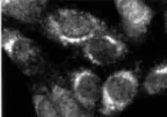
ICC/IF analysis of methanol-fixed HeLa cells using GTX23467 Furin antibody. Panel d represents the merged image showing cytoplasmic localization. Panel e shows the no primary antibody control. The images were captured at 60X magnification.
|

|
GTX23467 WB Image
WB analysis of various sample lysates using GTX23467 Furin antibody.
Dilution : 1:500
Loading : 30μg
|

|
GTX23467 WB Image
WB analysis of various sample lysates using GTX23467 Furin antibody. Expression of Furin was observed in HEK-293, HaCaT, BeWo, SK-O-V3, Mouse Liver and Rat Liver but not in Daudi and Ramos which are known to be low expressing cell lines (DOI: 10.1099/0022-1317-75-10-2821).
Dilution : 1:1000
Loading : 30μg
|

|
GTX23467 WB Image
WB analysis of HeLa (Lane 1), HeLa Cas9 (Lane 2), and HeLa Cas9 cells transduced with Furin Lentiviral sgRNA (Lane 3) cell lysates using GTX23467 Furin antibody(Fig. a). A loss of signal in sgRNA transduced cells using the LentiArray™ CRISPR product line confirms that the antibody is specific to Furin(Fig. b).
Dilution : 1:1000
Loading : 30μg
|

|
GTX23467 Image
ICC/IF analysis of 3T3 cells using GTX23467 Furin antibody.
|
REFERENCE
There are currently no references for Furin antibody (GTX23467). Be the first to share your publications with this product.
REVIEW
There are currently no reviews for Furin antibody (GTX23467). Be the first to share your experience with this product.


