AKT (phospho Ser473) antibody

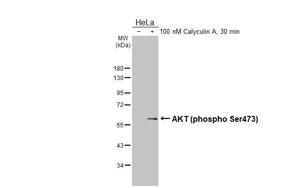
Untreated (–) and treated (+) HeLa whole cell extracts (30 μg) were separated by 10% SDS-PAGE, and the membrane was blotted with AKT (phospho Ser473) antibody (GTX128414) diluted at 1:500. The HRP-conjugated anti-rabbit IgG antibody (GTX213110-01) was used to detect the primary antibody.
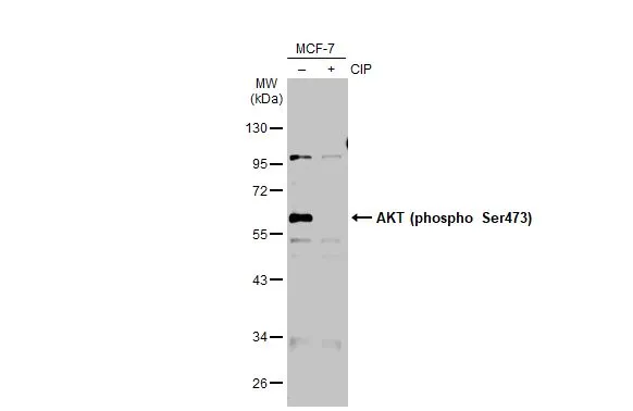
Untreated (–) and treated (+) MCF-7 whole cell extracts (30 μg) were separated by 10% SDS-PAGE, and the membrane was blotted with AKT (phospho Ser473) antibody (GTX128414) diluted at 1:500. The HRP-conjugated anti-rabbit IgG antibody (GTX213110-01) was used to detect the primary antibody.
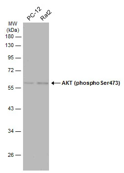
Various whole cell extracts (30 μg) were separated by 10% SDS-PAGE, and the membrane was blotted with AKT (phospho Ser473) antibody (GTX128414) diluted at 1:500. The HRP-conjugated anti-rabbit IgG antibody (GTX213110-01) was used to detect the primary antibody, and the signal was developed with Trident ECL plus-Enhanced.
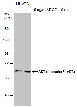
Untreated (–) and treated (+) HUVEC whole cell extracts (30 μg) were separated by 7.5% SDS-PAGE, and the membrane was blotted with AKT (phospho Ser473) antibody (GTX128414) diluted at 1:500. The HRP-conjugated anti-rabbit IgG antibody (GTX213110-01) was used to detect the primary antibody.
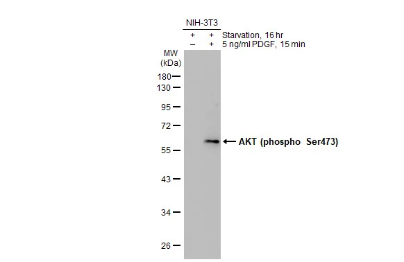
Untreated (–) and treated (+) NIH-3T3 whole cell extracts (30 μg) were separated by 10% SDS-PAGE, and the membrane was blotted with AKT (phospho Ser473) antibody (GTX128414) diluted at 1:500. The HRP-conjugated anti-rabbit IgG antibody (GTX213110-01) was used to detect the primary antibody.
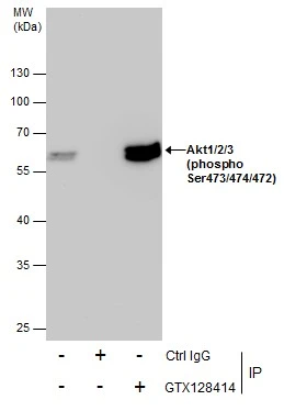
Immunoprecipitation of Akt1/2/3 (phospho Ser473/474/472) protein from 293T whole cell extracts using 5 μg of Akt1/2/3 (phospho Ser473/474/472) antibody (GTX128414).
Western blot analysis was performed using Akt1/2/3 (phospho Ser473/474/472) antibody (GTX128414).
EasyBlot anti-Rabbit IgG (GTX221666-01) was used as a secondary reagent.
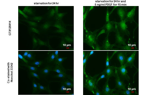
AKT (phospho Ser473) antibody detects AKT (phospho Ser473) protein by immunofluorescent analysis.Sample: NIH3T3 cells were fixed in 4% paraformaldehyde at RT for 15 min.Green: AKT (phospho Ser473) stained by AKT (phospho Ser473) antibody (GTX128414) diluted at 1:500.Blue: Hoechst 33342 staining.Scale bar= 10 μm.
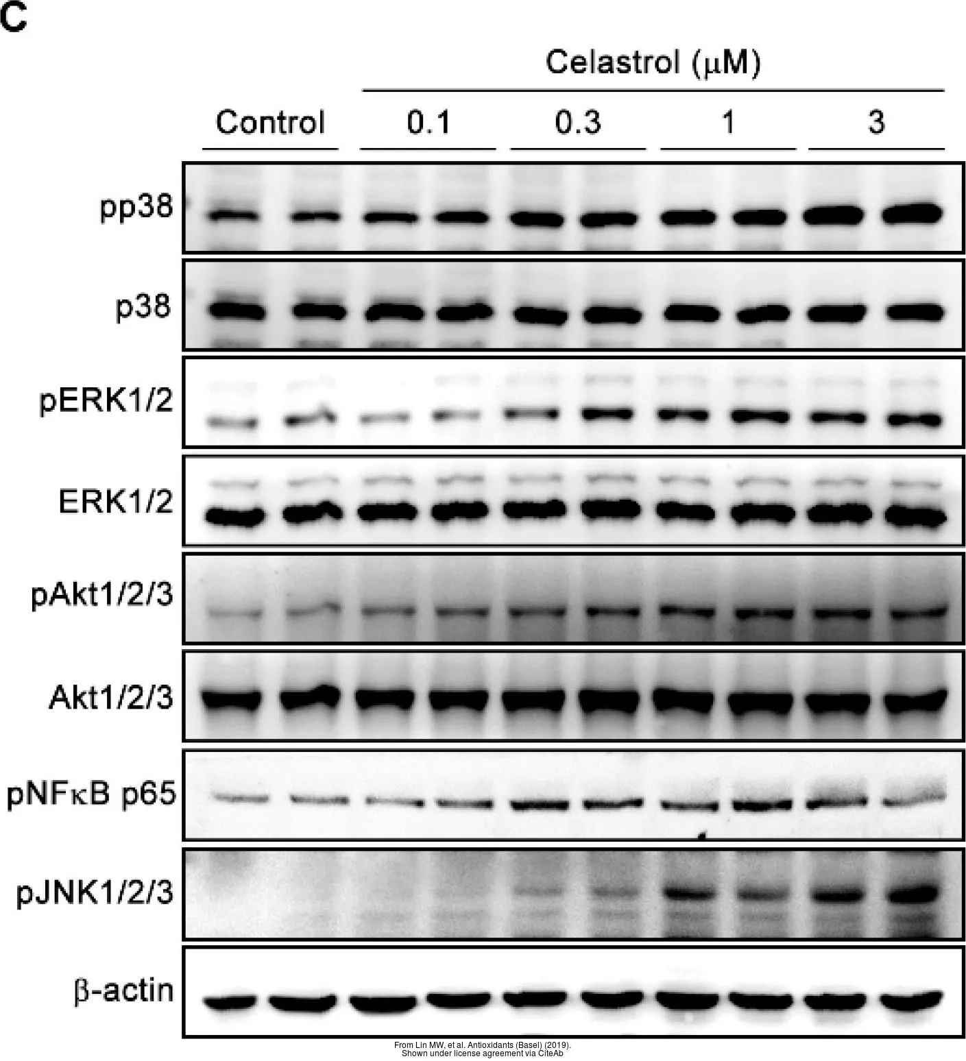
The data was published in the journal Antioxidants (Basel) in 2019.PMID: 31906147
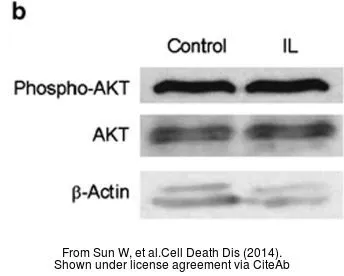
The data was published in the journal Cell Death Dis in 2014. PMID: 24457964
-
HostRabbit
-
ClonalityPolyclonal
-
IsotypeIgG
-
ApplicationsWB ICC/IF IHC-P IP
-
ReactivityHuman, Mouse, Rat, Pig