Actin Gamma 1 antibody
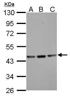
Sample (30 ug of whole cell lysate)
A: NIH-3T3
B: JC
C: BCL-1
10% SDS PAGE
GTX101794 diluted at 1:1000
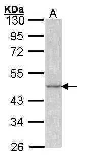
Sample (30 ug of whole cell lysate)
A: A431 (GTX27909)
10% SDS PAGE
GTX101794 diluted at 1:1000
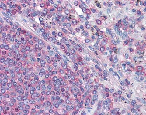
Immunohistochemical analysis of paraffin-embedded human Spleen, using gamma Actin(GTX101794) antibody(10 μg/ml).
Antigen Retrieval: Trilogy™ (EDTA based, pH 8.0) buffer, 15min
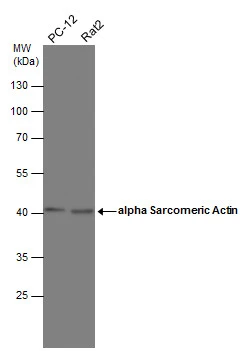
Various whole cell extracts (30 μg) were separated by 10% SDS-PAGE, and the membrane was blotted with alpha Sarcomeric Actin antibody (GTX101794) diluted at 1:1000.

Actin Gamma 1 antibody detects Actin Gamma 1 protein at cell membrane by immunohistochemical analysis.Sample: Paraffin-embedded cat intestine.Actin Gamma 1 stained by Actin Gamma 1 antibody (GTX101794) diluted at 1:500.Antigen Retrieval: Citrate buffer, pH 6.0, 15 min
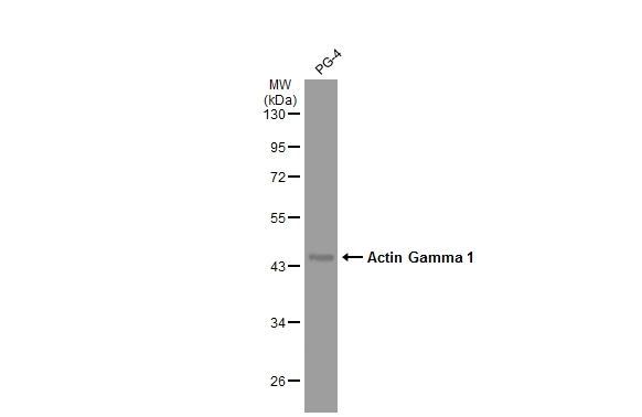
PG-4 whole cell extract (30 μg) was separated by 10% SDS-PAGE, and the membrane was blotted with Actin Gamma 1 antibody (GTX101794) diluted at 1:1000. The HRP-conjugated anti-rabbit IgG antibody (GTX213110-01) was used to detect the primary antibody.
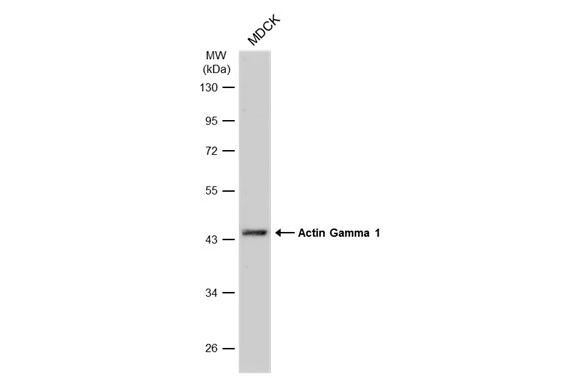
Whole cell extract (30 μg) was separated by 10% SDS-PAGE, and the membrane was blotted with Actin Gamma 1 antibody (GTX101794) diluted at 1:1000. The HRP-conjugated anti-rabbit IgG antibody (GTX213110-01) was used to detect the primary antibody, and the signal was developed with Trident ECL plus-Enhanced.
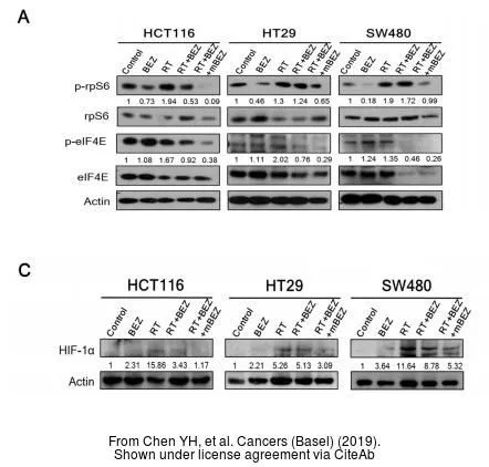
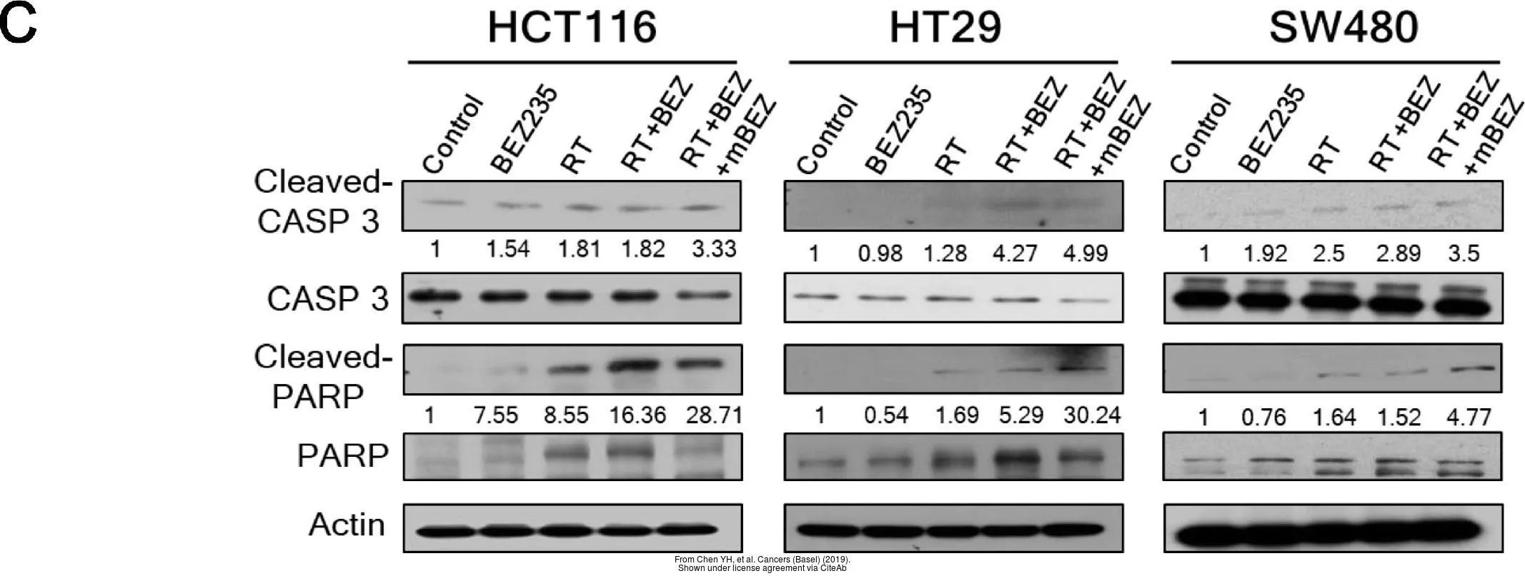
The data was published in the journal Cancers (Basel) in 2019.PMID: 31430901
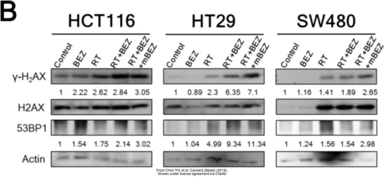
The data was published in the journal Cancers (Basel) in 2019.PMID: 31430901
The data was published in the journal Cancers (Basel) in 2019.PMID: 31430901
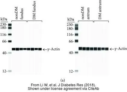
The data was published in the journal J Diabetes Res in 2018.PMID: 29955616
-
HostRabbit
-
ClonalityPolyclonal
-
IsotypeIgG
-
ApplicationsWB IHC-P IHC-Fr
-
ReactivityHuman, Mouse, Rat, Cat, Dog