BCL10 antibody


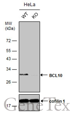
Wild-type (WT) and BCL10 knockout (KO) HeLa cell extracts (30 μg) were separated by 12% SDS-PAGE, and the membrane was blotted with BCL10 antibody (GTX112744) diluted at 1:2500. The HRP-conjugated anti-rabbit IgG antibody (GTX213110-01) was used to detect the primary antibody.
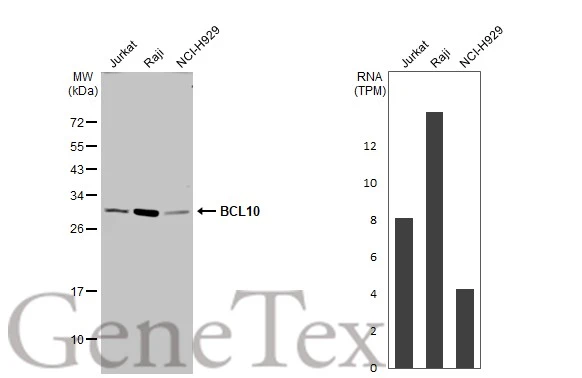
Various whole cell extracts (30 μg) were separated by 12% SDS-PAGE, and the membrane was blotted with BCL10 antibody (GTX112744) diluted at 1:1000. The HRP-conjugated anti-rabbit IgG antibody (GTX213110-01) was used to detect the primary antibody. Corresponding RNA expression data for the same cell lines are based on Human Protein Atlas program.
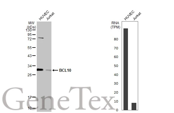
Various whole cell extracts (30 μg) were separated by 12% SDS-PAGE, and the membrane was blotted with BCL10 antibody (GTX112744) diluted at 1:1000. The HRP-conjugated anti-rabbit IgG antibody (GTX213110-01) was used to detect the primary antibody. Corresponding RNA expression data for the same cell lines are based on Human Protein Atlas program.
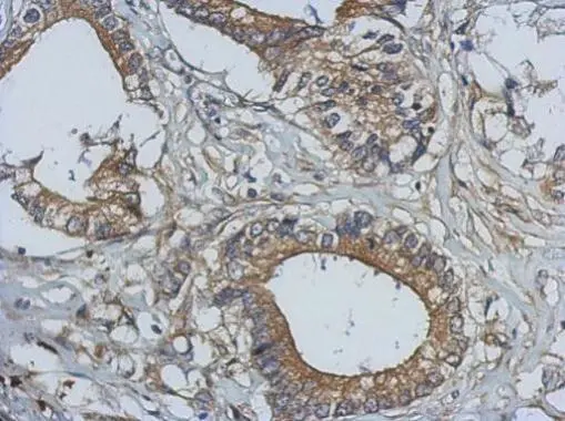
Immunohistochemical analysis of paraffin-embedded human gastric cancer, using BCL10 antibody(GTX112744) antibody at 1:500 dilution.
Antigen Retrieval: Trilogy™ (EDTA based, pH 8.0) buffer, 15min
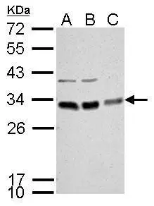
BCL10 antibody detects BCL10 protein by Western blot analysis.
A. 30 μg Neuro2A whole cell lysate/extract
B. 30 μg GL261 whole cell lysate/extract
C. 30 μg C8D30 whole cell lysate/extract
12 % SDS-PAGE
BCL10 antibody (GTX112744) dilution: 1:1000
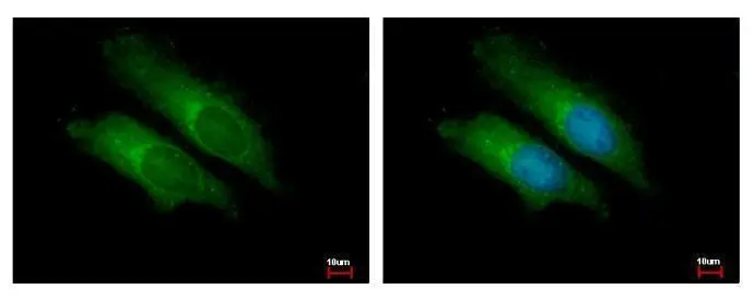
BCL10 antibody detects BCL10 protein at cytoplasm by immunofluorescent analysis.
Sample: HeLa cells were fixed in 2% paraformaldehyde/culture medium at 37ºC for 30 min.
Green: BCL10 protein stained by BCL10 antibody (GTX112744) diluted at 1:500.
Blue: Hoechst 33342 staining.
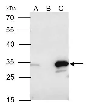
BCL10 antibody immunoprecipitates BCL10 protein in IP experiments.
IP samples: Raji whole cell extract
A. 40 μg Raji whole cell extract
B. Control with 4 μg of preimmune Rabbit IgG
C. Immunoprecipitation of BCL10 protein by 4 μg BCL10 antibody (GTX112744)
12 % SDS-PAGE
The immunoprecipitated BCL10 protein was detected by BCL10 antibody (GTX112744) diluted at 1:500.
[EasyBlot anti-rabbit IgG (GTX221666-01) was used as a secondary reagent]
-
HostRabbit
-
ClonalityPolyclonal
-
IsotypeIgG
-
ApplicationsWB ICC/IF IHC-P IP
-
ReactivityHuman, Mouse