BDNF antibody

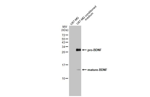
U87-MG whole cell extract and conditioned medium (30 μg) were separated by 15% SDS-PAGE, and the membrane was blotted with BDNF antibody (GTX132621) diluted at 1:125. The HRP-conjugated anti-rabbit IgG antibody (GTX213110-01) was used to detect the primary antibody.
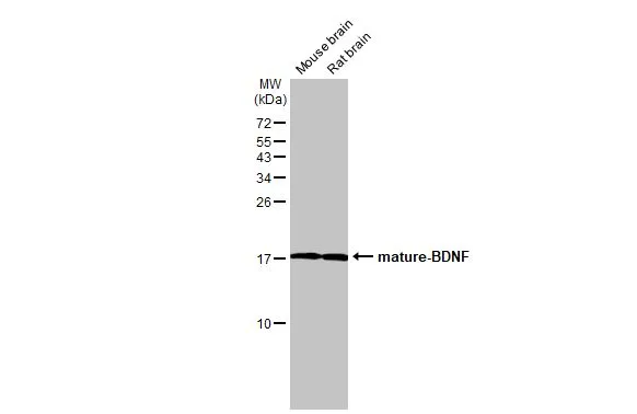
Various tissue extracts (50 μg) were separated by 15% SDS-PAGE, and the membrane was blotted with BDNF antibody (GTX132621) diluted at 1:500. The HRP-conjugated anti-rabbit IgG antibody (GTX213110-01) was used to detect the primary antibody.
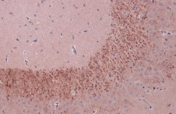
BDNF antibody detects BDNF protein at cytoplasm by immunohistochemical analysis.
Sample: Paraffin-embedded rat hippocampus CA3.
BDNF stained by BDNF antibody (GTX132621) diluted at 1:500.
Antigen Retrieval: Citrate buffer, pH 6.0, 15 min
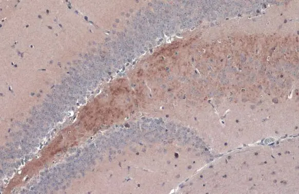
BDNF antibody detects BDNF protein at cytoplasm by immunohistochemical analysis.
Sample: Paraffin-embedded mouse hippocampus DG.
BDNF stained by BDNF antibody (GTX132621) diluted at 1:1000.
Antigen Retrieval: Citrate buffer, pH 6.0, 15 min
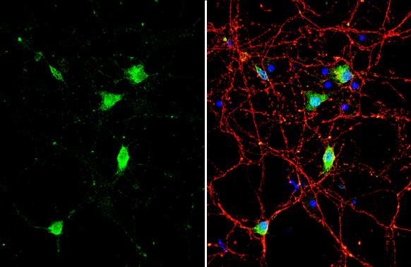
BDNF antibody detects BDNF protein by immunofluorescent analysis.Sample: DIV10 rat E18 primary cortical neuron cells were fixed in 4% paraformaldehyde at RT for 15 min.Green: BDNF stained by BDNF antibody (GTX132621) diluted at 1:500.Red: Tau, stained by Tau antibody [GT287] (GTX634809) diluted at 1:500.Blue: Fluoroshield with DAPI (GTX30920).
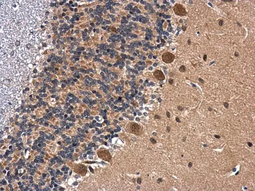
BDNF antibody detects BDNF protein at cytoplasm in rat brain by immunohistochemical analysis.
Sample: Paraffin-embedded rat brain.
BDNF antibody (GTX132621) diluted at 1:500.
Antigen Retrieval: Citrate buffer, pH 6.0, 15 min
-
HostRabbit
-
ClonalityPolyclonal
-
IsotypeIgG
-
ApplicationsWB ICC/IF IHC-P IHC-Fr
-
ReactivityHuman, Mouse, Rat