Bcl-X antibody

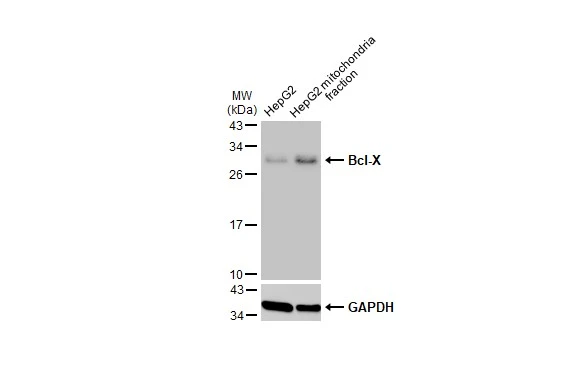
HepG2 and mitochondria extracts (30 μg) were separated by SDS-PAGE, and the membrane was blotted with Bcl-X antibody (GTX105661) diluted at 1:1000. The HRP-conjugated anti-rabbit IgG antibody (GTX213110-01) was used to detect the primary antibody.
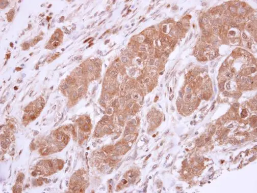
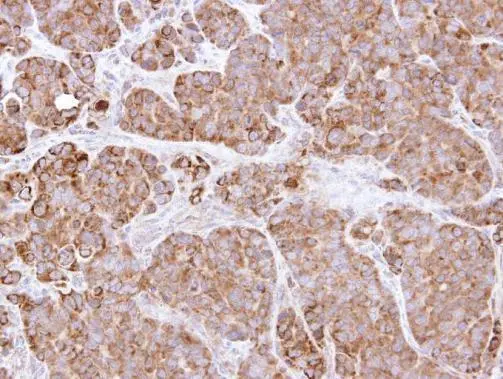
Bcl-X antibody detects Bcl-X protein at cytoplasm in human breast carcinoma by immunohistochemical analysis.
Sample: Paraffin-embedded human breast carcinoma.
Bcl-X antibody (GTX105661) diluted at 1:500.
Antigen Retrieval: Trilogy™ (EDTA based, pH 8.0) buffer, 15min
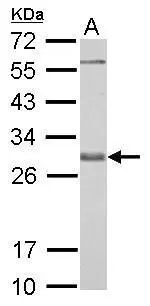
Bcl-X antibody detects BCL2L1 protein by western blot analysis.
A. 30 μg PC-12 whole cell lysate/extract
12% SDS-PAGE
Bcl-X antibody (GTX105661) dilution: 1:1000
The HRP-conjugated anti-rabbit IgG antibody (GTX213110-01) was used to detect the primary antibody.
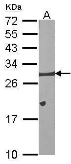
Sample (30 μg of whole cell lysate)
A:NIH-3T3
12% SDS PAGE
GTX105661 diluted at 1:1000
The HRP-conjugated anti-rabbit IgG antibody (GTX213110-01) was used to detect the primary antibody.
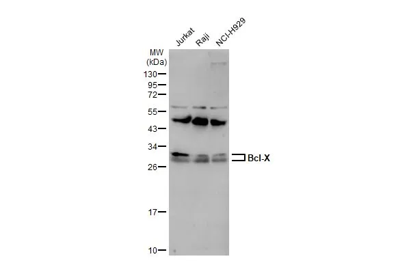
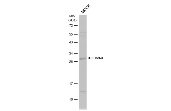
Various whole cell extracts (30 μg) were separated by 12% SDS-PAGE, and the membrane was blotted with Bcl-X antibody (GTX105661) diluted at 1:1000. The HRP-conjugated anti-rabbit IgG antibody (GTX213110-01) was used to detect the primary antibody.
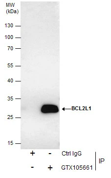
Immunoprecipitation of Bcl-X protein from HCT-116 whole cell extracts using 5 μg of Bcl-X antibody (GTX105661).
Western blot analysis was performed using Bcl-X antibody (GTX105661) diluted at 1:500.
EasyBlot anti-Rabbit IgG (GTX221666-01) was used as a secondary reagent.
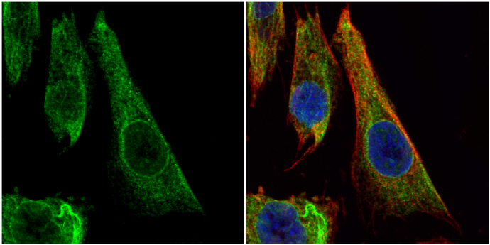
Bcl-X antibody detects Bcl-X protein at cytoplasm by immunofluorescent analysis.
Sample: SK-N-SH cells were fixed in 4% paraformaldehyde at RT for 15 min.
Green: Bcl-X protein stained by Bcl-X antibody (GTX105661) diluted at 1:200.
Red: alpha Tubulin, a cytoskeleton marker, stained by alpha Tubulin antibody [GT114] (GTX628802) diluted at 1:1000.
Blue: Hoechst 33342 staining.
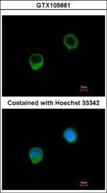
Immunofluorescence analysis of methanol-fixed A549, using BCL-x(GTX105661) antibody at 1:500 dilution.
Immunohistochemical analysis of paraffin-embedded SW480 xenograft, using BCL2L1(GTX105661) antibody at 1:500 dilution.
Antigen Retrieval: Trilogy™ (EDTA based, pH 8.0) buffer, 15min
Whole cell extract (30 μg) was separated by 12% SDS-PAGE, and the membrane was blotted with Bcl-X antibody (GTX105661) diluted at 1:1000. The HRP-conjugated anti-rabbit IgG antibody (GTX213110-01) was used to detect the primary antibody, and the signal was developed with Trident ECL plus-Enhanced.
-
HostRabbit
-
ClonalityPolyclonal
-
IsotypeIgG
-
ApplicationsWB ICC/IF IHC-P IP
-
ReactivityHuman, Mouse, Rat, Dog