Caveolin 2 antibody

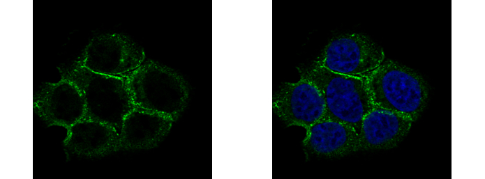
Caveolin 2 antibody detects Caveolin 2 protein at membrane by immunofluorescent analysis.
Sample: A431 cells were fixed in 4% paraformaldehyde at RT for 15 min.
Green: Caveolin 2 protein stained by Caveolin 2 antibody (GTX108294) diluted at 1:500.
Blue: Hoechst 33342 staining.
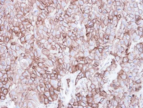
Immunohistochemical analysis of paraffin-embedded FaDu xenograft, using Caveolin 2(GTX108294) antibody at 1:500 dilution.
Antigen Retrieval: Trilogy™ (EDTA based, pH 8.0) buffer, 15min
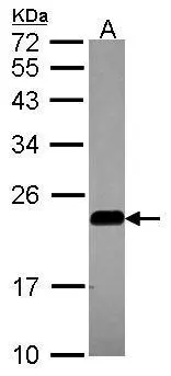
Sample (30 ug of whole cell lysate)
A: A549
12% SDS PAGE
GTX108294 diluted at 1:1000
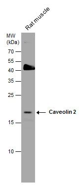
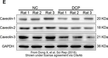
Caveolin 2 antibody detects Caveolin 2 protein by western blot analysis. Rat tissue extracts (50 μg) was separated by 15% SDS-PAGE, and the membrane was blotted with Caveolin 2 antibody (GTX108294) diluted at 1:1000.
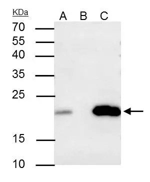
Caveolin 2 antibody immunoprecipitates caveolin 2 protein in IP experiments.
IP samples: A549 whole cell extract
A. 30 μg A549 whole cell extract
B. Control with 4 μg of preimmune Rabbit IgG
C. Immunoprecipitation of caveolin 2 protein by 4 μg Caveolin 2 antibody (GTX108294)
15 % SDS-PAGE
The immunoprecipitated caveolin 2 protein was detected by Caveolin 2 antibody (GTX108294) diluted at 1:500.
[EasyBlot anti-rabbit IgG (GTX221666-01) was used as a secondary reagent]
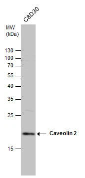
Caveolin 2 antibody detects Caveolin 2 protein by western blot analysis. Whole cell extracts (30 μg) was separated by 15% SDS-PAGE, and the membrane was blotted with Caveolin 2 antibody (GTX108294) diluted at 1:1000.
-
HostRabbit
-
ClonalityPolyclonal
-
IsotypeIgG
-
ApplicationsWB ICC/IF IHC-P IP
-
ReactivityHuman, Mouse, Rat