Collagen III antibody
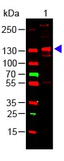
WB analysis of human collagen III protein using GTX27778 Collagen III antibody.
Loading : 100 ng
Dilution : 1:1000
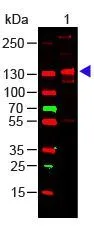
Western Blot of Rabbit Anti-Collagen III Antibody (GTX27778) Load: 100 ng Human Collagen III. Primary antibody: Collagen III Antibody at 1:1000 o/n at 4ºC. Secondary antibody: DyLight™ 649 Goat anti-rabbit at 1:20,000 for 30 min at RT Block for 30 min at RT. Predicted/Observed size: 138 kDa, 138 kDa.
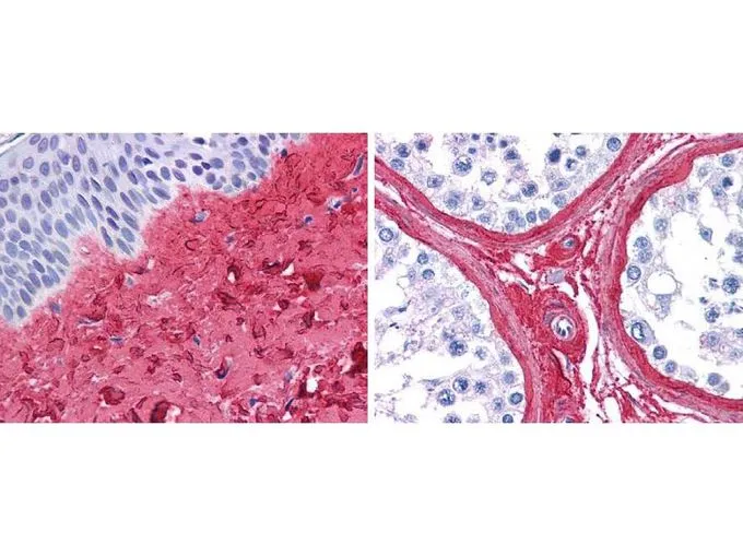
GeneTex anti collagen III antibody (GTX27778, 45 min RT) showed strong staining in FFPE sections of human skin(left, dermis) with moderate to strong red staining and testis (right) where strong staining was observed within connective tissue between seminiferous tubules. The antibody showed strong extracellular staining within connective tissues across many organs with minimal background staining. Slides were steamed in 0.01 M sodium citrate buffer, pH 6.0 at 99-100ºC - 20 minutes for antigen retrieval.
The data was published in the journal Oncotarget in 2017. PMID: 28465479
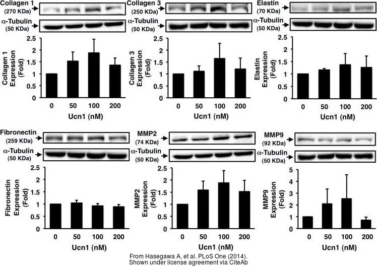
The data was published in the journal PLoS One in 2014. PMID: 25462164
-
HostRabbit
-
ClonalityPolyclonal
-
IsotypeIgG
-
ApplicationsWB ICC/IF IHC-P IHC-Fr FCM IP Dot ELISA Multiplexing
-
ReactivityHuman, Mouse, Bovine, Pig