Cyclin T1 antibody
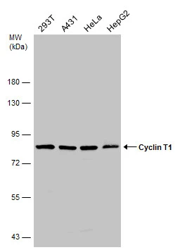
Various whole cell extracts (50 μg) were separated by 7.5% SDS-PAGE, and the membrane was blotted with Cyclin T1 antibody (GTX133413) diluted at 1:1000.
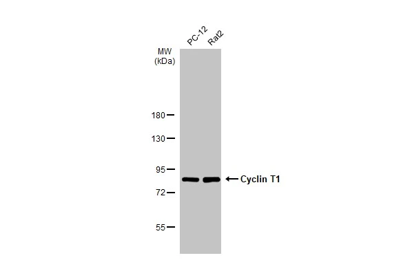
Various whole cell extracts (30 μg) were separated by 7.5% SDS-PAGE, and the membrane was blotted with Cyclin T1 antibody (GTX133413) diluted at 1:1000. The HRP-conjugated anti-rabbit IgG antibody (GTX213110-01) was used to detect the primary antibody.
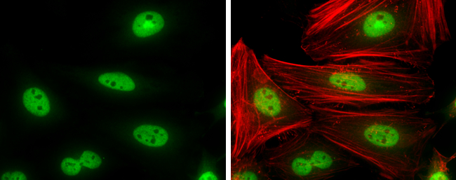
Cyclin T1 antibody detects Cyclin T1 protein at nucleus by immunofluorescent analysis.
Sample: HeLa cells were fixed in 4% paraformaldehyde at RT for 15 min.
Green: Cyclin T1 protein stained by Cyclin T1 antibody (GTX133413) diluted at 1:500.
Red: Phalloidin, a cytoskeleton marker, diluted at 1:100.
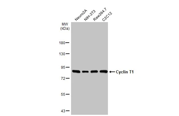
Various whole cell extracts (30 μg) were separated by 7.5% SDS-PAGE, and the membrane was blotted with Cyclin T1 antibody (GTX133413) diluted at 1:1000. The HRP-conjugated anti-rabbit IgG antibody (GTX213110-01) was used to detect the primary antibody.
-
HostRabbit
-
ClonalityPolyclonal
-
IsotypeIgG
-
ApplicationsWB ICC/IF
-
ReactivityHuman, Mouse, Rat