DC-SIGN antibody
Cat. No. GTX31656
Cat. No. GTX31656
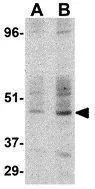
GTX31656 WB Image
WB analysis of human small intestine tissue lysate using GTX31656 DC-SIGN antibody.
Working concentration : (A) 1 and (B) 2 μg/ml
1 / 3
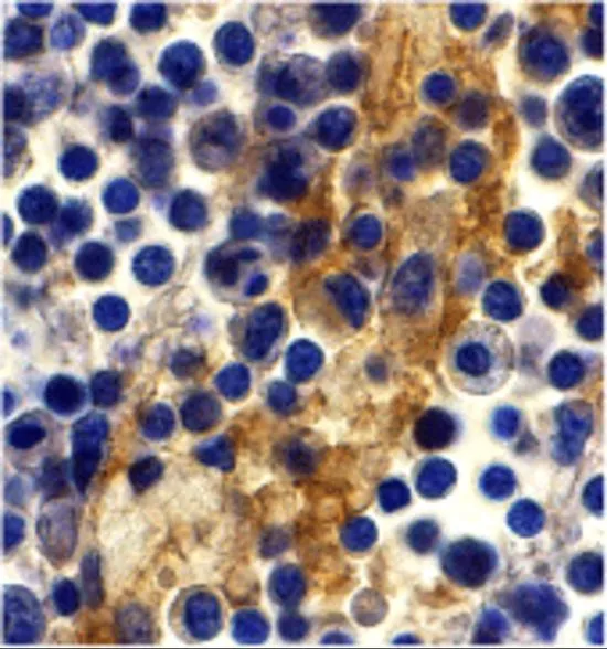
GTX31656 IHC-P Image
IHC-P analysis of human lymph node tissue using GTX31656 DC-SIGN antibody.
Working concentration : 10 μg/ml
2 / 3
GTX31656 IHC-P Image
IHC-P analysis of human lymph node tissue using GTX31656 DC-SIGN antibody.
Working concentration : 20 μg/ml
3 / 3
-
HostRabbit
-
ClonalityPolyclonal
-
IsotypeIgG
-
ApplicationsWB IHC-P ELISA
-
ReactivityHuman