ER81 antibody


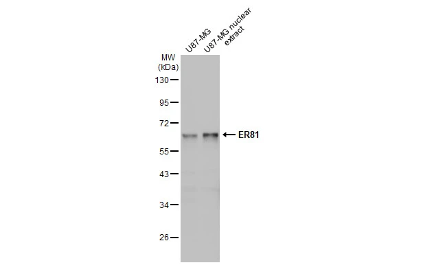
U87-MG whole cell and nuclear extracts (30 μg) were separated by 10% SDS-PAGE, and the membrane was blotted with ER81 antibody (GTX129202) diluted at 1:1000. The HRP-conjugated anti-rabbit IgG antibody (GTX213110-01) was used to detect the primary antibody.
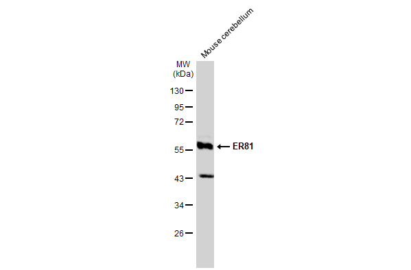
Mouse tissue extract (50 μg) was separated by 10% SDS-PAGE, and the membrane was blotted with ER81 antibody (GTX129202) diluted at 1:1000. The HRP-conjugated anti-rabbit IgG antibody (GTX213110-01) was used to detect the primary antibody.
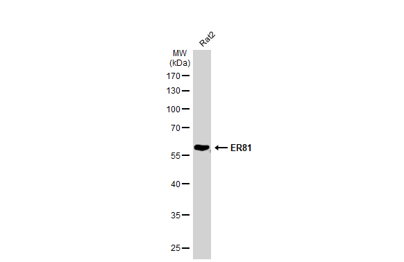
Whole cell extract (30 μg) was separated by 10% SDS-PAGE, and the membrane was blotted with ER81 antibody (GTX129202) diluted at 1:1000. The HRP-conjugated anti-rabbit IgG antibody (GTX213110-01) was used to detect the primary antibody.
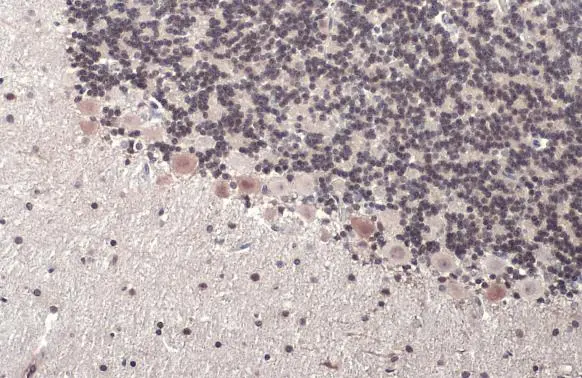
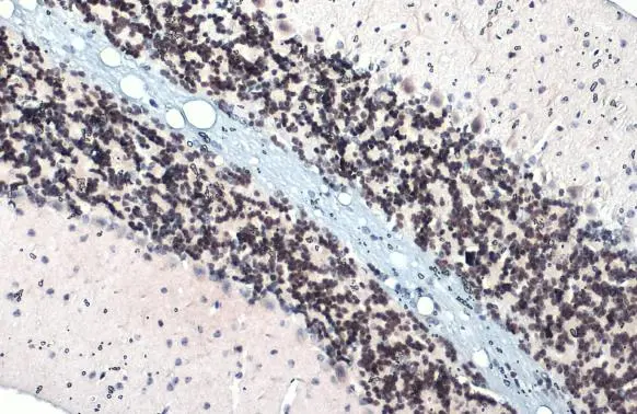
ER81 antibody detects ER81 protein at nucleus by immunohistochemical analysis.Sample: Paraffin-embedded rat brain.ER81 stained by ER81 antibody (GTX129202) diluted at 1:500.Antigen Retrieval: Citrate buffer, pH 6.0, 15 min
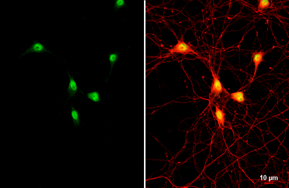
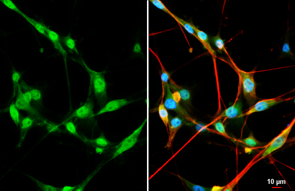
ER81 antibody detects ER81 protein by immunofluorescent analysis.Sample: DIV9 rat hippocampal neuron and Glia cell cells were fixed in 4% paraformaldehyde at RT for 15 min.Green: ER81 stained by ER81 antibody (GTX129202) diluted at 1:250.Red: Tau, an axon marker, stained by Tau antibody [GT287] (GTX634809) diluted at 1:500.
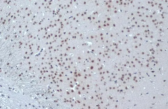
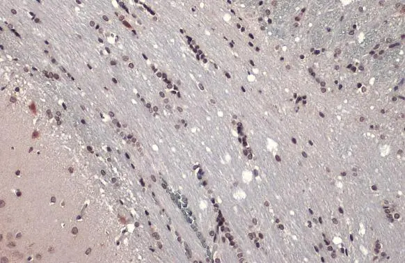
ER81 antibody detects ER81 protein at nucleus by immunohistochemical analysis.Sample: Paraffin-embedded mouse brain.ER81 stained by ER81 antibody (GTX129202) diluted at 1:500.Antigen Retrieval: Citrate buffer, pH 6.0, 15 min
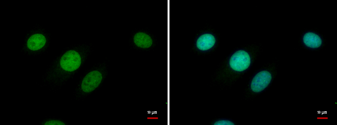
ER81 antibody detects ER81 protein at nucleus by immunofluorescent analysis.
Sample: C8D30 cells were fixed in 4% paraformaldehyde at RT for 15 min.
Green: ER81 protein stained by ER81 antibody (GTX129202) diluted at 1:500.
Blue: Hoechst 33342 staining.
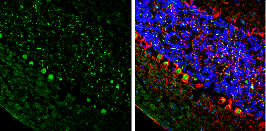
ER81 antibody detects ER81 protein by immunohistochemical analysis.Sample: Frozen-sectioned mouse cerebellum.Green: ER81 stained by ER81 antibody (GTX129202) diluted at 1:250.Red: NF-H, stained by NF-H antibody [GT114] (GTX634289) diluted at 1:500.Blue: Fluoroshield with DAPI (GTX30920).
ER81 antibody detects ER81 protein at nucleus by immunohistochemical analysis.Sample: Paraffin-embedded mouse brain.ER81 stained by ER81 antibody (GTX129202) diluted at 1:500.Antigen Retrieval: Citrate buffer, pH 6.0, 15 min
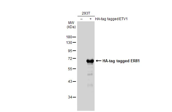
Non-transfected (–) and transfected (+) 293T whole cell extracts (30 μg) were separated by 10% SDS-PAGE, and the membrane was blotted with ER81 antibody (GTX129202) diluted at 1:5000. The HRP-conjugated anti-rabbit IgG antibody (GTX213110-01) was used to detect the primary antibody.
ER81 antibody detects ER81 protein at nucleus by immunohistochemical analysis.Sample: Paraffin-embedded rat brain.ER81 stained by ER81 antibody (GTX129202) diluted at 1:500.Antigen Retrieval: Citrate buffer, pH 6.0, 15 min
ER81 antibody detects ER81 protein at nucleus by immunofluorescent analysis.Sample: U87-MG cells were fixed in 4% paraformaldehyde at RT for 15 min.Green: ER81 stained by ER81 antibody (GTX129202) diluted at 1:200.Red: alpha Tubulin, a cytoskeleton marker, stained by alpha Tubulin antibody [GT114] (GTX628802) diluted at 1:1000.Blue: Fluoroshield with DAPI (GTX30920).Scale bar= 10μm.
-
HostRabbit
-
ClonalityPolyclonal
-
IsotypeIgG
-
ApplicationsWB ICC/IF IHC-P IHC-Fr
-
ReactivityHuman, Mouse, Rat