GAD67 antibody

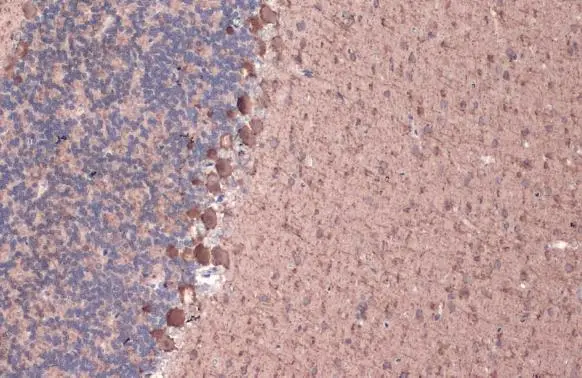
GAD67 antibody detects GAD67 protein at cell membrane and cytoplasm by immunohistochemical analysis.Sample: Paraffin-embedded rat cerebellum.GAD67 stained by GAD67 antibody (GTX101881) diluted at 1:1000.Antigen Retrieval: Citrate buffer, pH 6.0, 15 min
GAD67 antibody detects GAD67 protein at cell membrane and cytoplasm by immunohistochemical analysis.Sample: Paraffin-embedded mouse cerebellum.GAD67 stained by GAD67 antibody (GTX101881) diluted at 1:1000.Antigen Retrieval: Citrate buffer, pH 6.0, 15 min
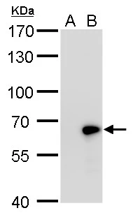
GAD67 antibody detects GAD67 protein by western blot analysis.
A. 30 μg 293T whole cell lysate/extract
B. 30 μg whole cell lysate/extract of human GAD1-transfected 293T cells
7.5% SDS-PAGE
GAD67 antibody (GTX101881) dilution: 1:5000
The HRP-conjugated anti-rabbit IgG antibody (GTX213110-01) was used to detect the primary antibody.
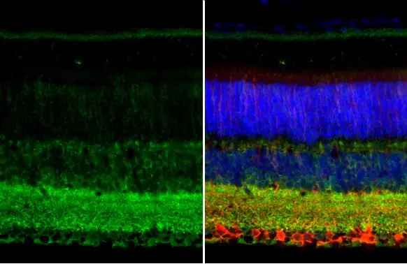
GAD67 antibody detects GAD67 protein at cell membrane and cytoplasm by immunohistochemical analysis.Sample: Paraffin-embedded mouse eye.Green: GAD67 stained by GAD67 antibody (GTX101881) diluted at 1:250.Red: beta Tubulin 3/ Tuj1, a neural marker, stained by beta Tubulin 3/ Tuj1 antibody [GT11710] (GTX631836) diluted at 1:500.Blue: Fluoroshield with DAPI (GTX30920).Antigen Retrieval: Citrate buffer, pH 6.0, 15 min
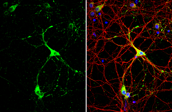
GAD67 antibody detects GAD67 protein by immunofluorescent analysis.Sample: DIV10 rat E18 primary cortical neuron cells were fixed in 4% paraformaldehyde at RT for 15 min.Green: GAD67 stained by GAD67 antibody (GTX101881) diluted at 1:500.Red: Tau, stained by Tau antibody [GT287] (GTX634809) diluted at 1:500.Blue: Fluoroshield with DAPI (GTX30920).
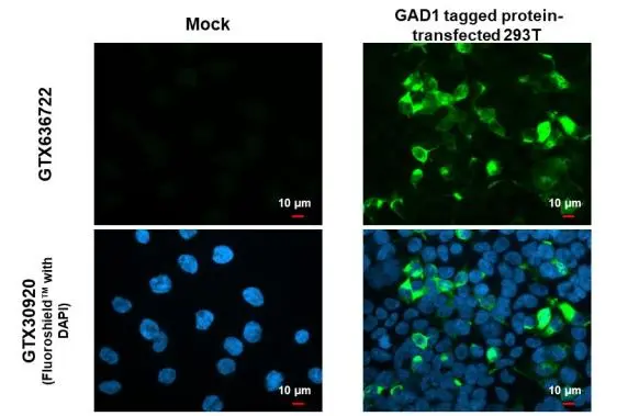
GAD67 antibody detects GAD67 protein by immunofluorescent analysis.Sample: Mock and transfected 293T cells were fixed in 4% paraformaldehyde at RT for 15 min.Green: GAD67 stained by GAD67 antibody (GTX101881) diluted at 1:500.Blue: Fluoroshield with DAPI (GTX30920).
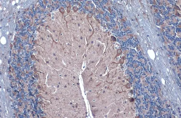
GAD67 antibody detects GAD67 protein at cytoplasm by immunohistochemical analysis.Sample: Paraffin-embedded mouse brain.GAD67 stained by GAD67 antibody (GTX101881) diluted at 1:500.Antigen Retrieval: Citrate buffer, pH 6.0, 15 min
Various whole cell extracts (30 μg) were separated by 7.5% SDS-PAGE, and the membrane was blotted with GAD67 antibody (GTX101881) diluted at 1:500. The HRP-conjugated anti-rabbit IgG antibody (GTX213110-01) was used to detect the primary antibody.
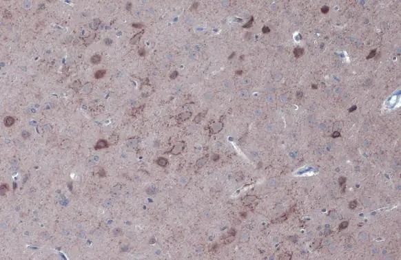
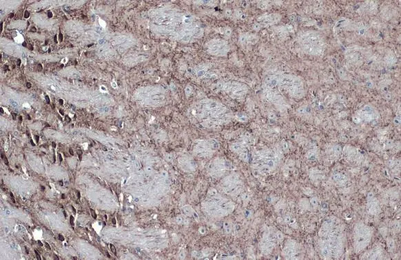
GAD67 antibody detects GAD67 protein at cytoplasm by immunohistochemical analysis.Sample: Paraffin-embedded rat brain.GAD67 stained by GAD67 antibody (GTX101881) diluted at 1:500.Antigen Retrieval: Citrate buffer, pH 6.0, 15 min
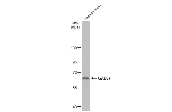
Human tissue extract (30 μg) was separated by 7.5% SDS-PAGE, and the membrane was blotted with GAD67 antibody (GTX101881) diluted at 1:1000. The HRP-conjugated anti-rabbit IgG antibody (GTX213110-01) was used to detect the primary antibody.
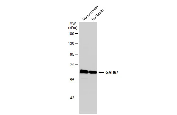
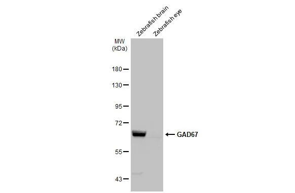
Various tissue extracts (50 μg) were separated by 7.5% SDS-PAGE, and the membrane was blotted with GAD67 antibody (GTX101881) diluted at 1:1000. The HRP-conjugated anti-rabbit IgG antibody (GTX213110-01) was used to detect the primary antibody.
GAD67 antibody detects GAD67 protein on myofiber membranes by immunohistochemical analysis.Sample: Whole-mount zebrafish embryo GAD67 antibody (GTX101881) dilution: 1:200.
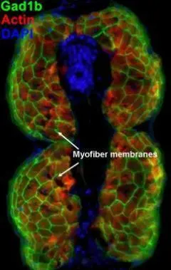
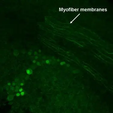
GAD67 antibody detects GAD67 protein on myofiber membranes by immunohistochemical analysis.Sample: Agarose-embedded zebrafish embryo GAD67 antibody (GTX101881) dilution: 1:200.Image provided with permission courtesy of Dr. T. Schilling at UC, Irvine.
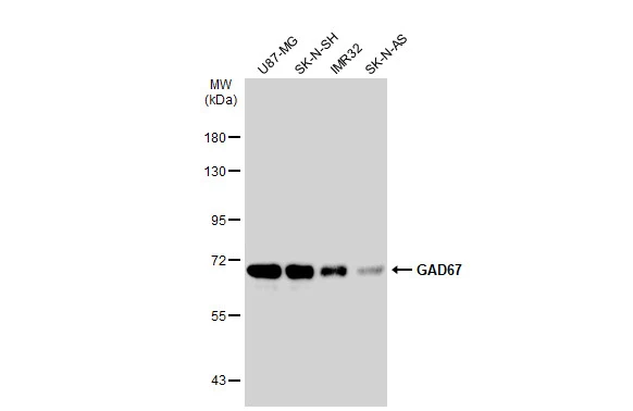
Various tissue extracts (30 μg) were separated by 7.5% SDS-PAGE, and the membrane was blotted with GAD67 antibody (GTX101881) diluted at 1:500. The HRP-conjugated anti-rabbit IgG antibody (GTX213110-01) was used to detect the primary antibody.
-
HostRabbit
-
ClonalityPolyclonal
-
IsotypeIgG
-
ApplicationsWB ICC/IF IHC-P IHC-Fr IHC-Wm IHC IHC (Free Floating)
-
ReactivityHuman, Mouse, Rat, Zebrafish, Monkey