GEF-H1 antibody

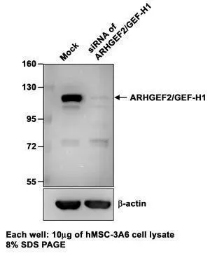
Sample (10 μg of whole cell lysate)
A: hMSC-3A6
B: siRNA of ARHGEF2/GEF-H1
8% SDS PAGE
GTX125893 diluted at 1:1000
The HRP-conjugated anti-rabbit IgG antibody (GTX213110-01) was used to detect the primary antibody.
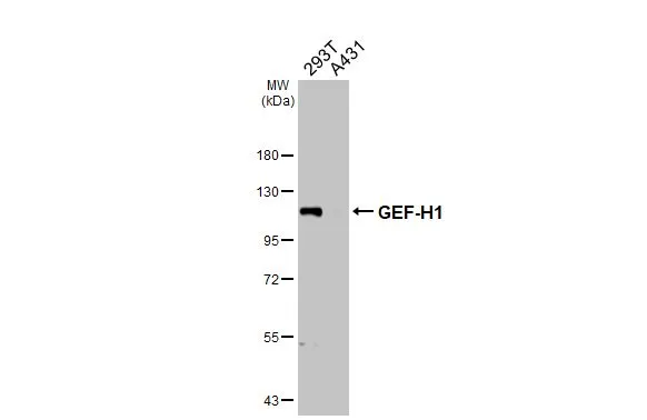
Various whole cell extracts (30 μg) were separated by 7.5% SDS-PAGE, and the membrane was blotted with GEF-H1 antibody (GTX125893) diluted at 1:2000. The HRP-conjugated anti-rabbit IgG antibody (GTX213110-01) was used to detect the primary antibody.
GEF-H1 antibody detects GEF-H1 protein at cytoplasm by immunohistochemical analysis.Sample: Paraffin-embedded mouse brain.GEF-H1 stained by GEF-H1 antibody (GTX125893) diluted at 1:1000.Antigen Retrieval: Citrate buffer, pH 6.0, 15 min
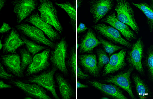
GEF-H1 antibody detects GEF-H1 protein at cytoskeleton and Golgi apparatus by immunofluorescent analysis.Sample: HeLa cells were fixed in ice-cold MeOH for 5 min.Green: GEF-H1 stained by GEF-H1 antibody (GTX125893) diluted at 1:500.Blue: Hoechst 33342 staining.Scale bar= 10 μm.
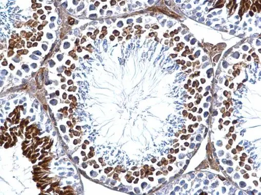
GEF-H1 antibody detects GEF-H1 protein at cytosol on mouse testis by immunohistochemical analysis.
Sample: Paraffin-embedded mouse testis.
GEF-H1 antibody (GTX125893) dilution: 1:500.
Antigen Retrieval: Trilogy™ (EDTA based, pH 8.0) buffer, 15min
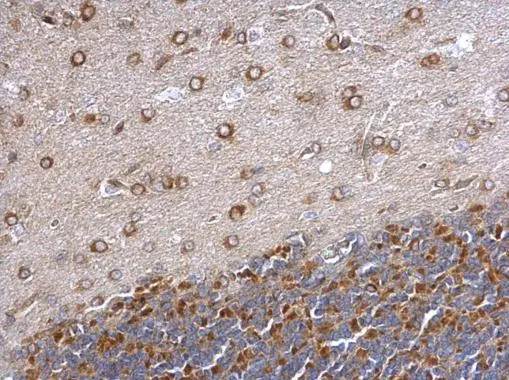
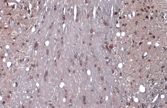
GEF-H1 antibody detects GEF-H1 protein at cytosol on rat hind brain by immunohistochemical analysis.
Sample: Paraffin-embedded rat hind brain.
GEF-H1 antibody (GTX125893) dilution: 1:500.
Antigen Retrieval: Trilogy™ (EDTA based, pH 8.0) buffer, 15min
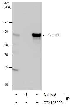
Immunoprecipitation of GEF-H1 protein from 293T whole cell extracts using 5 μg of GEF-H1 antibody (GTX125893).
Western blot analysis was performed using GEF-H1 antibody (GTX125893).
EasyBlot anti-Rabbit IgG (GTX221666-01) was used as a secondary reagent.
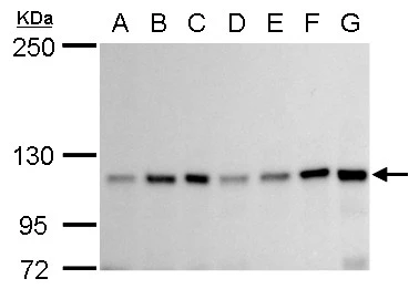
GEF-H1 antibody detects GEF-H1 protein by western blot analysis.
A. 30 μg Neuro2A whole cell lysate/extract
B. 30 μg GL261 whole cell lysate/extract
C. 30 μg C8D30 whole cell lysate/extract
D. 30 μg NIH-3T3 whole cell lysate/extract
E. 30 μg BCL-1 whole cell lysate/extract
F. 30 μg Raw 264.7 whole cell lysate/extract
G. 30 μg C2Cl2 whole cell lysate/extract
5% SDS-PAGE
GEF-H1 antibody (GTX125893) dilution: 1:1000
The HRP-conjugated anti-rabbit IgG antibody (GTX213110-01) was used to detect the primary antibody.
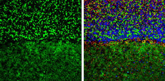
GEF-H1 antibody detects GEF-H1 protein by immunohistochemical analysis.Sample: Frozen-sectioned mouse mouse cerebellum.Green: GEF-H1 stained by GEF-H1 antibody (GTX125893) diluted at 1:250.Red: NF-H, stained by NF-H antibody [GT114] (GTX634289) diluted at 1:500.Blue: Fluoroshield with DAPI (GTX30920).
Antigen Retrieval: Citrate buffer, pH 6.0, 10 min
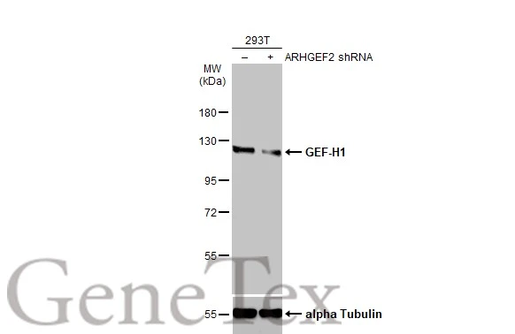
Non-transfected (–) and transfected (+) 293T whole cell extracts (30 μg) were separated by 7.5% SDS-PAGE, and the membrane was blotted with GEF-H1 antibody (GTX125893) diluted at 1:4000. The HRP-conjugated anti-rabbit IgG antibody (GTX213110-01) was used to detect the primary antibody.
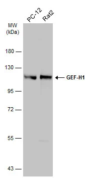
Various whole cell extracts (30 μg) were separated by 7.5% SDS-PAGE, and the membrane was blotted with GEF-H1 antibody (GTX125893) diluted at 1:500. The HRP-conjugated anti-rabbit IgG antibody (GTX213110-01) was used to detect the primary antibody.
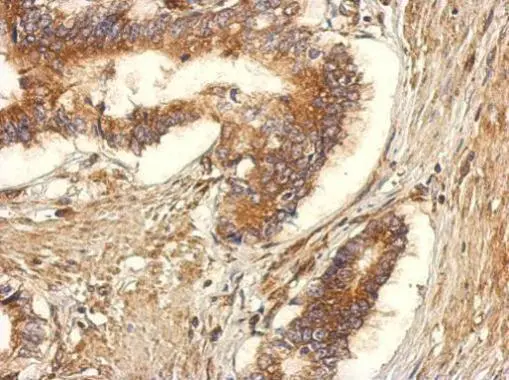
Immunohistochemical analysis of paraffin-embedded human gastric cancer, using GEF-H1(GTX125893) antibody at 1:500 dilution.
Antigen Retrieval: Trilogy™ (EDTA based, pH 8.0) buffer, 15min
-
HostRabbit
-
ClonalityPolyclonal
-
IsotypeIgG
-
ApplicationsWB ICC/IF IHC-P IHC-Fr IP
-
ReactivityHuman, Mouse, Rat