GFP antibody
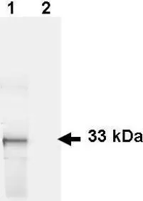
Figure 1. Western blot of GFP recombinant protein detected with GeneTex polyclonal anti-GFP antibody. Lane 1 shows detection of a 33 kDa band corresponding to a GFP containing recombinant protein (arrowhead) expressed in HeLa cells. Lane 2 shows no staining of a mock transfected HeLa cell lysate. After blocking the membrane was probed with the primary antibody diluted to 1 μg/ml for 1 h at room temperature followed by washes and reaction with a 1:2,500 dilution of donkey anti-Goat IgG [H&L].
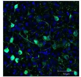
Sf-1+ neurons and their processes of the ventromedial nucleus of the hypothalamus in Mus musculus (coronal view, 20X magnification). GeneTex polyclonal goat anti-GFP was used at 1:500 dilution in free floating imunnohistochemistry to detect eGFP. Fluorchrome conjugated anti-goat IgG secondary antibody was used for detection at 1:500. Sections were counterstained with DAPI. Personal Communication, Daniel A. Lee, Johns Hopkins School of Medicine, Baltimore, MD.
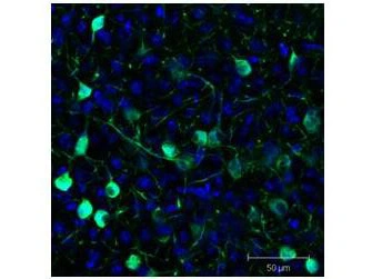
IHC (free floating) analysis of Sf-1:Cre mice crossed to the Z/EG reporter line, Mouse brain using GTX26673 GFP antibody.
Green : Primary antibody
Blue : DAPI
Dilution : 1:500
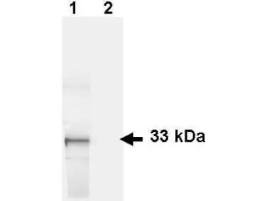
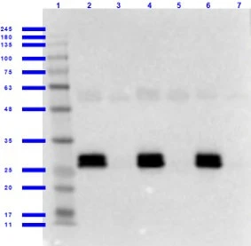
WB analysis of various samples using GTX26673 GFP antibody.
Lane 1 : GFP-transfected HeLa whole cell lysate
Lane 2 : HeLa whole cell lysate
Loading : 35 μg
Dilution : 1 μg/mL
WB analysis of various samples using GTX26673 GFP antibody.
Lane 1 : Protein ladder
Lane 2 : Human serum + GFP
Lane 3 : Human serum only
Lane 4 : Mouse serum + GFP
Lane 5 : Mouse serum only
Lane 6 : Rat serum + GFP
Lane 7 : Rat serum only
Dilution : 1 μg/mL
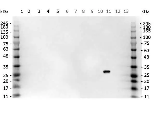
WB analysis of various samples using GTX26673 GFP antibody.
Lane 1 : 293T whole cell lysate
Lane 2 : HeLa whole cell lysate
Lane 3 : CHO-K1 whole cell lysate
Lane 4 : MDA-MB-231 whole cell lysate
Lane 5 : A431 whole cell lysate
Lane 6 : Jurkat whole cell lysate
Lane 7 : NIH-3T3 whole cell lysate
Lane 8 : HCP
Lane 9 : Flag-tagged protein
Lane 10 : RFP
Lane 11 : GFP
Lane 12 : GST-tagged protein
Lane 13 : MBP
Dilution : 1 μg/mL
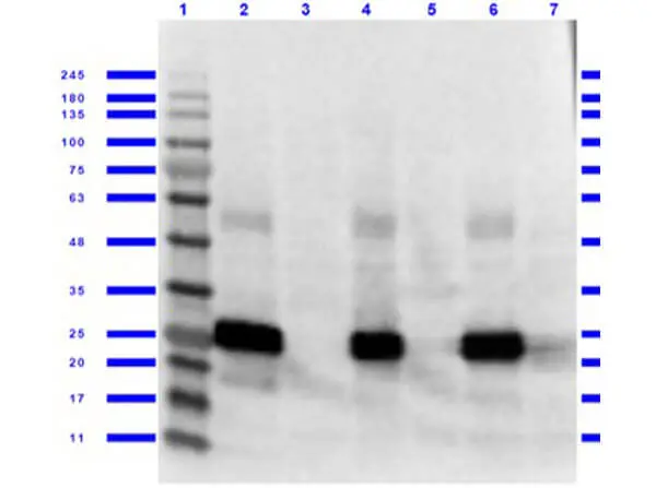
WB analysis of various samples using GTX26673 GFP antibody.
Lane 1 : Protein ladder
Lane 2 : 293T cells+ GFP
Lane 3 : 293T cells only
Lane 4 : NIH-3T3 cells + GFP
Lane 5 : NIH-3T3 cells only
Lane 6 : PC-12 cells + GFP
Lane 7 : PC-12 cells only
Dilution : 1 μg/mL

ICC/IF analysis of GFP transgenic mouse embryo cells using GTX26673 GFP antibody.
Green : Primary antibody
Blue : DAPI
Dilution :1:500
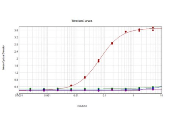
ELISA analysis of various samples using serially diluted GTX26673 GFP antibody.
Red : GF'P
Green : Human IgG
Blue : Mouse IgG
Purple : Rat IgG
Coating : 1 μg
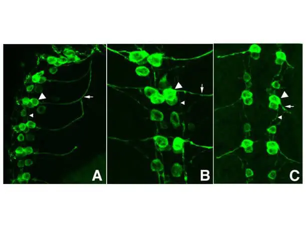
ICC/IF analysis of PFA-fixed Drosophila melanogaster late stage embryonic central nervous using GTX26673 GFP antibody.
Panel A : shows a lateral view (ventral left)
Panels B and C : shows ventral views of whole mount embryos at 63x magnification (plus 2x digital zoom).
In all panels, anterior is up. Tau-GFP cell bodies (large arrowhead) and axons of motorneurons (arrow) and interneurons (small arrowhead) as green fluorescent signal.
Dillution : 1:1000
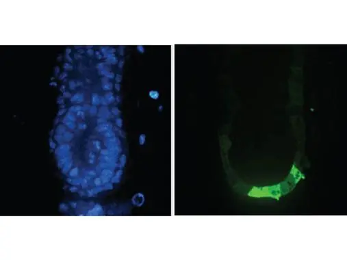
Tissue: E5.5 Hex-GFP transgenic mouse embryo. Primary antibody: Goat anti-GFP was used at 1:500 dilution. Secondary antibody: Fluorchrome conjugated Anti-goat IgG secondary antibody at 1:10,000 for 45 min at RT. Staining: GFP as green fluorescent signal with DAPI blue counterstain.
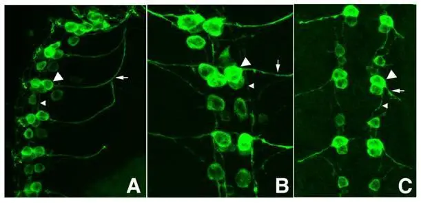
Figure 2. Immunofluorescence microscopy. GeneTex polyclonal anti-GFP antibody at a 1:1,000 dilution detects tau-GFP in cell bodies (large arrowhead) and axons of motorneurons (arrow) and interneurons (small arrowhead) in Drosophila melanogaster late stage embryonic central nervous system. Fluorochrome conjugated anti-Goat secondary antibody was used for detection at 1:300. Panel A shows a lateral view (ventral left) and Panels B and C show ventral views of whole mount embryos at 63x magnification (plus 2x digital zoom). Personal Communication, Helmata Mistry, Washington University School of Medicine, St. Louis, MO.
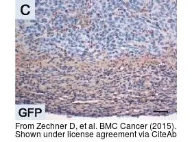
The data was published in the journal BMC Cancer in 2015. PMID: 25885700
-
HostGoat
-
ClonalityPolyclonal
-
IsotypeIgG
-
ApplicationsWB ICC/IF IHC-P IHC-Fr FCM IP ELISA IHC Multiplexing Purification IHC (Free Floating)
-
ReactivitySpecies independent