Glutamine synthetase antibody


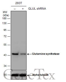
Non-transfected (–) and transfected (+) 293T whole cell extracts (30 μg) were separated by 10% SDS-PAGE, and the membrane was blotted with Glutamine synthetase antibody (GTX109121) diluted at 1:1000. The HRP-conjugated anti-rabbit IgG antibody (GTX213110-01) was used to detect the primary antibody.
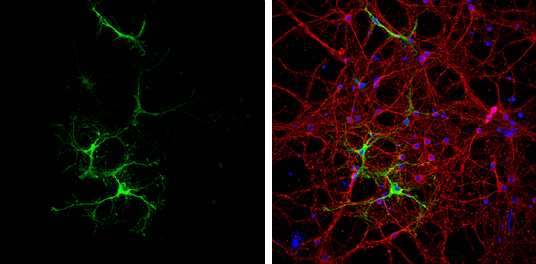
Glutamine synthetase antibody detects Glutamine synthetase protein at astrocytes by immunofluorescent analysis.
Sample: DIV9 rat E18 primary cortical neurons were fixed in 4% paraformaldehyde at RT for 15 min.
Green: Glutamine synthetase protein stained by Glutamine synthetase antibody (GTX109121) diluted at 1:500.
Red: beta Tubulin 3/ Tuj1, a neuron cell marker, stained by beta Tubulin 3/ Tuj1 antibody [GT11710] (GTX631836) diluted at 1:500.
Blue: Fluoroshield with DAPI (GTX30920).
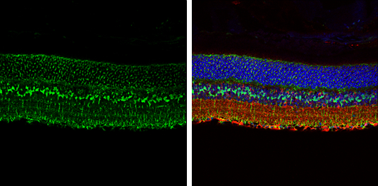
Glutamine synthetase antibody detects Glutamine synthetase protein expression by immunohistochemical analysis.
Sample:Paraffin-embedded adult mouse retina.
Green: Glutamine synthetase protein stained by Glutamine synthetase antibody (GTX109121) diluted at 1:250.
Red: beta Tubulin 3/ TUJ1, stained by beta Tubulin 3/ TUJ1 antibody [GT11710] (GTX631836) diluted at 1:250.
Blue: Fluoroshield with DAPI (GTX30920).
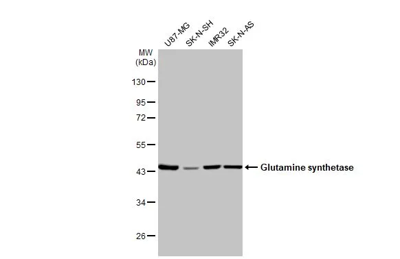
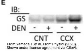
Various whole cell extracts (30 μg) were separated by 10% SDS-PAGE, and the membrane was blotted with Glutamine synthetase antibody (GTX109121) diluted at 1:1000. The HRP-conjugated anti-rabbit IgG antibody (GTX213110-01) was used to detect the primary antibody.
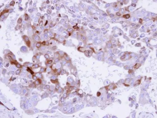
Immunohistochemical analysis of paraffin-embedded H441 xenograft , using Glutamine Synthetase (GTX109121) antibody at 1:500 dilution.
Antigen Retrieval: Trilogy™ (EDTA based, pH 8.0) buffer, 15min
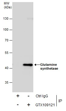
Immunoprecipitation of Glutamine synthetase protein from IMR32 whole cell extracts using 5 μg of Glutamine synthetase antibody (GTX109121).
Western blot analysis was performed using Glutamine synthetase antibody (GTX109121).
EasyBlot anti-Rabbit IgG (GTX221666-01) was used as a secondary reagent.
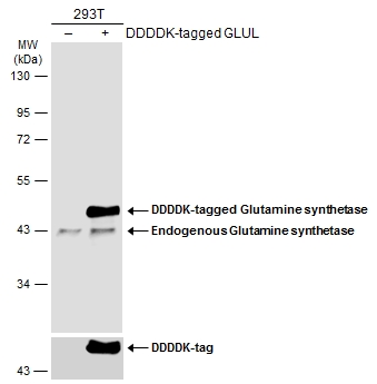
Non-transfected (–) and transfected (+) 293T whole cell extracts (30 μg) were separated by 10% SDS-PAGE, and the membrane was blotted with Glutamine synthetase antibody (GTX109121) diluted at 1:10000. The HRP-conjugated anti-rabbit IgG antibody (GTX213110-01) was used to detect the primary antibody.
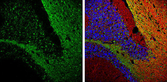
Glutamine synthetase antibody detects Glutamine synthetase protein expression by immunohistochemical analysis.
Sample: Frozen-sectioned adult mouse cerebellum.
Green: Glutamine synthetase protein stained by Glutamine synthetase antibody (GTX109121) diluted at 1:250.
Red: beta Tubulin 3/ TUJ1, stained by beta Tubulin 3/ TUJ1 antibody [GT11710] (GTX631836) diluted at 1:500.
Blue: Fluoroshield with DAPI (GTX30920).
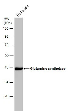
Rat tissue extract (50 μg) was separated by 10% SDS-PAGE, and the membrane was blotted with Glutamine synthetase antibody (GTX109121) diluted at 1:50000.
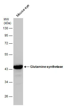
Mouse tissue extract (50 μg) was separated by 10% SDS-PAGE, and the membrane was blotted with Glutamine synthetase antibody (GTX109121) diluted at 1:20000.
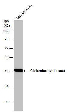
Mouse tissue extract (50 μg) was separated by 10% SDS-PAGE, and the membrane was blotted with Glutamine synthetase antibody (GTX109121) diluted at 1:50000.
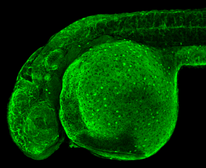
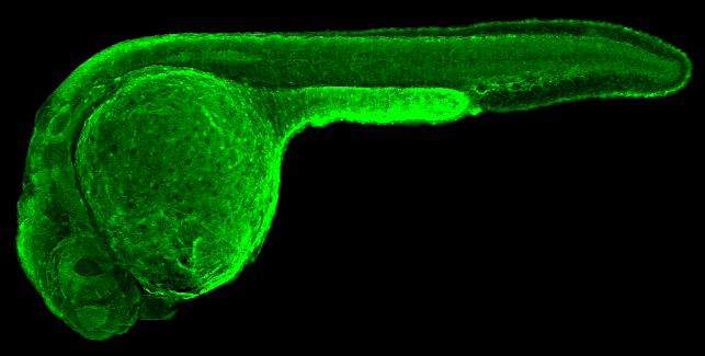
Glutamine synthetase antibody detects Glutamine synthetase protein on zebrafish by whole mount immunohistochemical analysis.
Sample: 2 days-post-fertilization zebrafish embryo.
Glutamine synthetase antibody (GTX109121) dilution: 1:100.
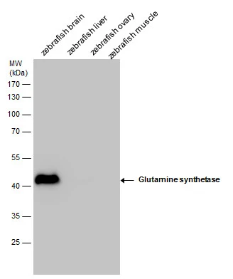
Glutamine synthetase antibody detects Glutamine synthetase protein by Western blot analysis. Zebrafish tissue extracts (30 μg) was separated by 10 % SDS-PAGE, and the membrane was blotted with Glutamine synthetase antibody (GTX109121) at a dilution of 1:10000.
Glutamine synthetase antibody detects Glutamine synthetase protein on zebrafish by whole mount immunohistochemical analysis.
Sample: 1 day-post-fertilization zebrafish embryo.
Glutamine synthetase antibody (GTX109121) dilution: 1:100.
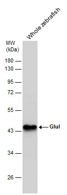
Whole zebrafish extract (30 μg) was separated by 10% SDS-PAGE, and the membrane was blotted with Glutamine synthetase antibody (GTX109121) diluted at 1:10000.
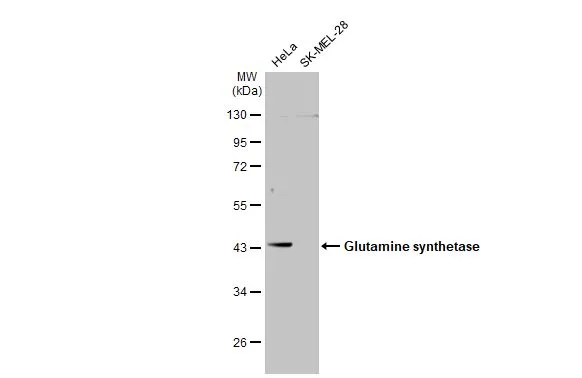
Various whole cell extracts (30 μg) were separated by 10% SDS-PAGE, and the membrane was blotted with Glutamine synthetase antibody (GTX109121) diluted at 1:1000. The HRP-conjugated anti-rabbit IgG antibody (GTX213110-01) was used to detect the primary antibody.
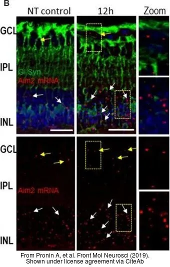
The data was published in the journal Front Mol Neurosci in 2019. PMID: 30930743
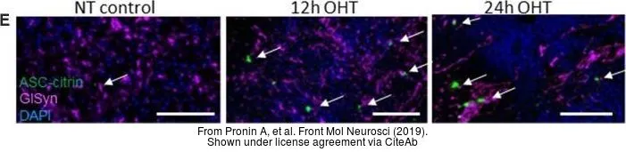
The data was published in the journal Front Mol Neurosci in 2019. PMID: 30930743
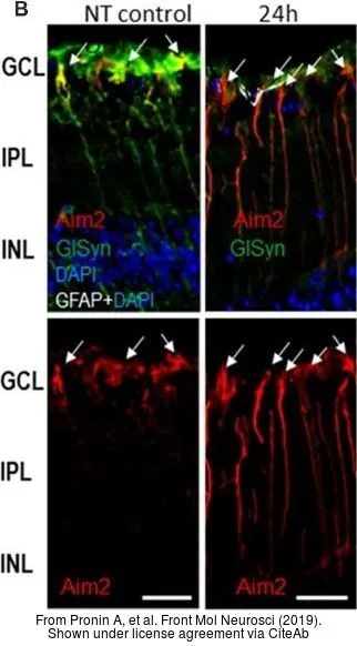
The data was published in the journal Front Mol Neurosci in 2019. PMID: 30930743
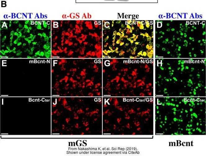
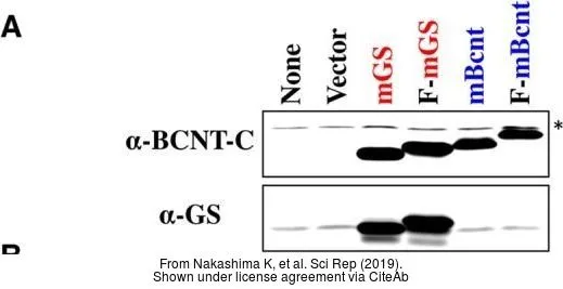
The data was published in the journal Sci Rep in 2019. PMID: 31616018
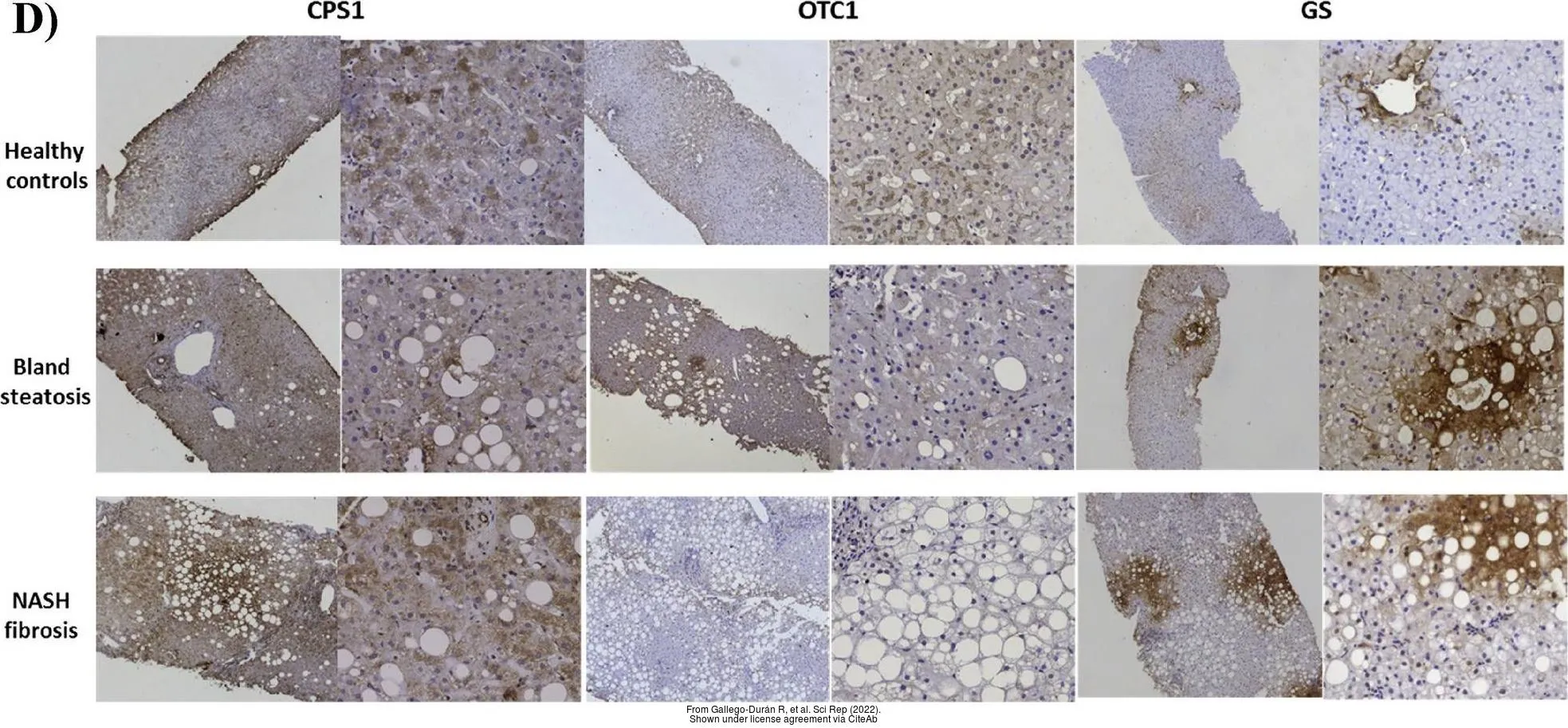
The data was published in the 2022 in Sci Rep. PMID: 35232986
-
HostRabbit
-
ClonalityPolyclonal
-
IsotypeIgG
-
ApplicationsWB ICC/IF IHC-P IHC-Fr IHC-Wm IP
-
ReactivityHuman, Mouse, Rat, Zebrafish