HDAC3 antibody

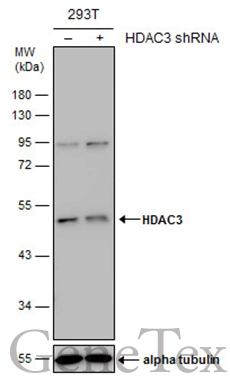
Non-transfected (–) and transfected (+) 293T whole cell extracts (30 μg) were separated by 10% SDS-PAGE, and the membrane was blotted with HDAC3 antibody (GTX113303) diluted at 1:500. The HRP-conjugated anti-rabbit IgG antibody (GTX213110-01) was used to detect the primary antibody.
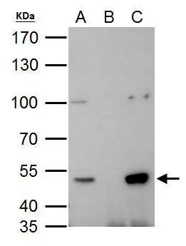
HDAC3 antibody immunoprecipitates HDAC3 protein in IP experiments. IP Sample: 293T whole cell lysate/extract A. 40 μg 293T whole cell lysate/extract B. Control with 2 μg of preimmune rabbit IgG C. Immunoprecipitation of HDAC3 protein by 2 μg of HDAC3 antibody (GTX113303) 7.5% SDS-PAGE The immunoprecipitated HDAC3 protein was detected by HDAC3 antibody (GTX113303) diluted at 1:1000. EasyBlot anti-rabbit IgG (GTX221666-01) was used as a secondary reagent.
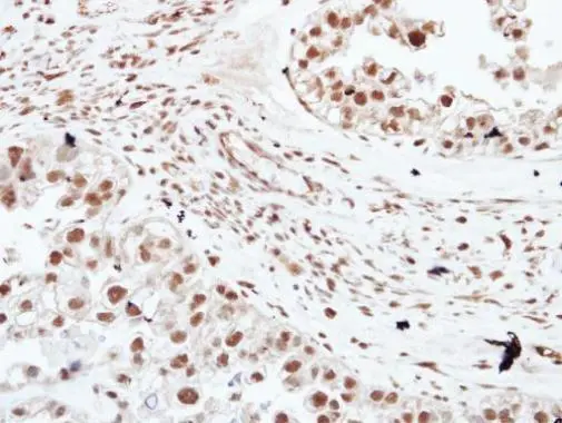
Immunohistochemical analysis of paraffin-embedded human ovarian cancer, using HDAC3(GTX113303) antibody at 1:250 dilution.
Antigen Retrieval: Citrate buffer, pH 6.0, 15 min
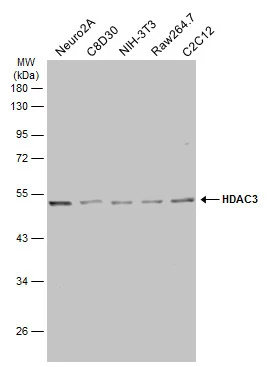
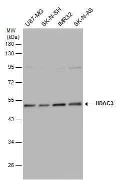
Various whole cell extracts (30 μg) were separated by 10% SDS-PAGE, and the membrane was blotted with HDAC3 antibody (GTX113303) diluted at 1:1000. The HRP-conjugated anti-rabbit IgG antibody (GTX213110-01) was used to detect the primary antibody.
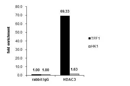
ChIP experiment and primer designs are based on Proc Natl Acad Sci U S A. 2005 Aug 16;102(33):11651-6.
Cross-linked ChIP was performed with MCF-7 chromatin extract and 5 μg of either control rabbit IgG or anti-HDAC3 antibody. The precipitated DNA was detected by PCR with primer set targeting to TFF1 or HK1.
Various whole cell extracts (30 μg) were separated by 10% SDS-PAGE, and the membrane was blotted with HDAC3 antibody (GTX113303) diluted at 1:1000. The HRP-conjugated anti-rabbit IgG antibody (GTX213110-01) was used to detect the primary antibody.
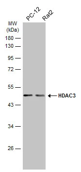
Various whole cell extracts (30 μg) were separated by 10% SDS-PAGE, and the membrane was blotted with HDAC3 antibody (GTX113303) diluted at 1:500. The HRP-conjugated anti-rabbit IgG antibody (GTX213110-01) was used to detect the primary antibody.
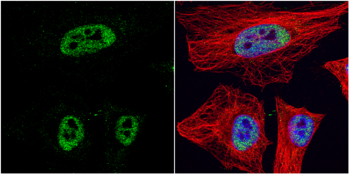
HDAC3 antibody detects HDAC3 protein at nucleus by immunofluorescent analysis.
Sample: HeLa cells were fixed in 4% paraformaldehyde at RT for 15 min.
Green: HDAC3 protein stained by HDAC3 antibody (GTX113303) diluted at 1:1000.
Red: alpha Tubulin, a cytoskeleton marker, stained by alpha Tubulin antibody [GT114] (GTX628802) diluted at 1:1000.
Blue: Hoechst 33342 staining.
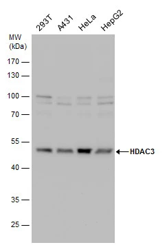
HDAC3 antibody detects HDAC3 protein by Western blot analysis. Various whole cell extracts (30 μg) were separated by 10% SDS-PAGE, and the membrane was blotted with HDAC3 antibody (GTX113303) diluted by 1:1000.
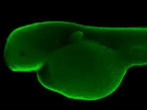
Immunohistochemical analysis (whole mount) of zebrafish embryo, using HDAC3 antibody (GTX113303) at 1:200 dilution.
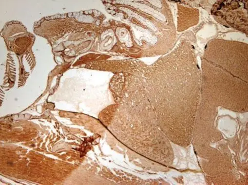
Immunohistochemical analysis of paraffin-embedded zebrafish tissue, using HDAC3 antibody (GTX113303) at 1:300 dilution.
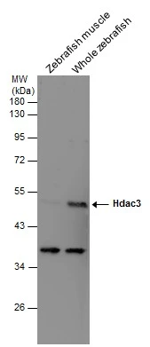
Various tissue extracts (30 μg) were separated by 10% SDS-PAGE, and the membrane was blotted with HDAC3 antibody (GTX113303) diluted at 1:500.
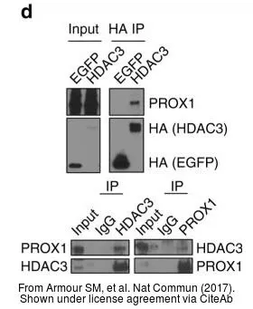
The data was published in the journal Nat Commun in 2017. PMID: 28916805
-
HostRabbit
-
ClonalityPolyclonal
-
IsotypeIgG
-
ApplicationsWB ICC/IF IHC-P IHC-Wm IP ChIP assay
-
ReactivityHuman, Mouse, Rat, Zebrafish