Histone H1.0 antibody

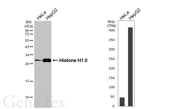
Various whole cell extracts (30 μg) were separated by 12% SDS-PAGE, and the membrane was blotted with Histone H1.0 antibody (GTX114462) diluted at 1:10000. The HRP-conjugated anti-rabbit IgG antibody (GTX213110-01) was used to detect the primary antibody. Corresponding RNA expression data for the same cell lines are based on Human Protein Atlas program.
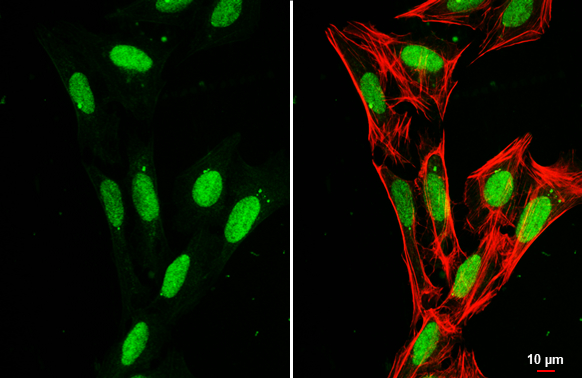
Histone H1.0 antibody detects Histone H1.0 protein at nucleus by immunofluorescent analysis.Sample: U2OS cells were fixed in 4% paraformaldehyde at RT for 15 min.Green: Histone H1.0 stained by Histone H1.0 antibody (GTX114462) diluted at 1:1000.Red: phalloidin, a cytoskeleton marker, diluted at 1:200.Scale bar= 10 μm.
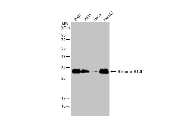
Various whole cell extracts (30 μg) were separated by 12% SDS-PAGE, and the membrane was blotted with Histone H1.0 antibody (GTX114462) diluted at 1:10000. The HRP-conjugated anti-rabbit IgG antibody (GTX213110-01) was used to detect the primary antibody.
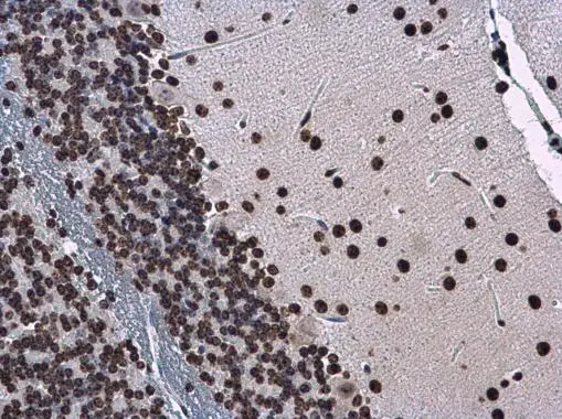
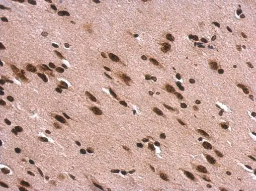
Histone H1.0 antibody detects Histone H1.0 protein at nucleus in mouse brain by immunohistochemical analysis.
Sample: Paraffin-embedded mouse brain.
Histone H1.0 antibody (GTX114462) diluted at 1:500.
Antigen Retrieval: Citrate buffer, pH 6.0, 15 min
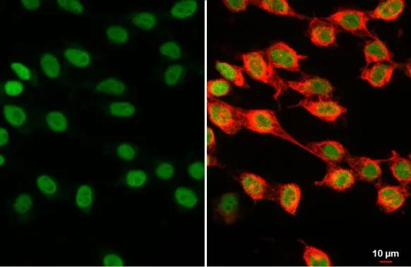
Histone H1.0 antibody detects Histone H1.0 protein at nucleus by immunofluorescent analysis.Sample: 293T cells were fixed in 4% paraformaldehyde at RT for 15 min.Green: Histone H1.0 stained by Histone H1.0 antibody (GTX114462) diluted at 1:500.Red: alpha Tubulin, a cytoskeleton marker, stained by alpha Tubulin antibody [GT114] (GTX628802) diluted at 1:1000.
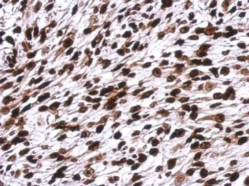
Immunohistochemical analysis of paraffin-embedded C2C12 xenograft, using Histone H1.0(GTX114462) antibody at 1:500 dilution.
Antigen Retrieval: Trilogy™ (EDTA based, pH 8.0) buffer, 15min
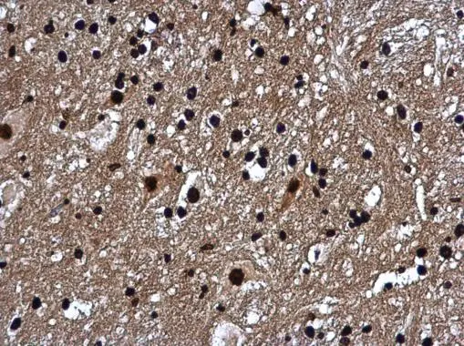
Histone H1.0 antibody detects Histone H1.0 protein at nucleus in mouse brain by immunohistochemical analysis.
Sample: Paraffin-embedded mouse brain.
Histone H1.0 antibody (GTX114462) diluted at 1:500.
Antigen Retrieval: Citrate buffer, pH 6.0, 15 min
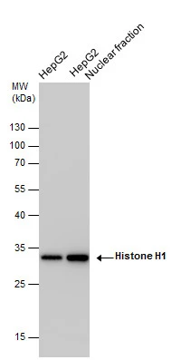
Histone H1 antibody detects Histone H1 protein by western blot analysis. HepG2 whole cell extracts and nuclear extracts (30 μg) were separated by 12% SDS-PAGE, and the membrane was blotted with Histone H1 antibody (GTX114462) at a dilution of 1:10000 and developed with Trident femto Western HRP Substrate (GTX14698). The HRP-conjugated anti-rabbit IgG antibody (GTX213110-01) was used to detect the primary antibody.
Histone H1.0 antibody detects Histone H1.0 protein at nucleus on rat fore brain by immunohistochemical analysis.
Sample: Paraffin-embedded rat fore brain.
Histone H1.0 antibody (GTX114462) dilution: 1:500.
Antigen Retrieval: Trilogy™ (EDTA based, pH 8.0) buffer, 15min
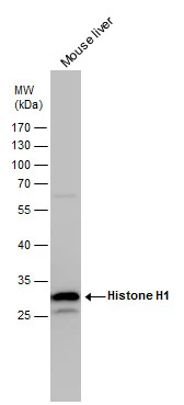
Histone H1 antibody detects Histone H1 protein by western blot analysis. Mouse tissue extracts (50 μg) was separated by 12% SDS-PAGE, and the membrane was blotted with Histone H1 antibody (GTX114462) diluted by 1:5000. The HRP-conjugated anti-rabbit IgG antibody (GTX213110-01) was used to detect the primary antibody.
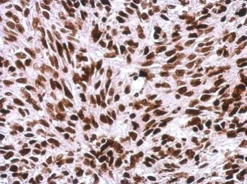
Immunohistochemical analysis of paraffin-embedded SkHep1 xenograft, using Histone H1.0(GTX114462) antibody at 1:500 dilution.
Antigen Retrieval: Trilogy™ (EDTA based, pH 8.0) buffer, 15min
-
HostRabbit
-
ClonalityPolyclonal
-
IsotypeIgG
-
ApplicationsWB ICC/IF IHC-P
-
ReactivityHuman, Mouse, Rat