IDH1 antibody


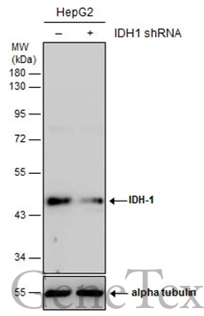
Non-transfected (–) and transfected (+) HepG2 whole cell extracts (30 μg) were separated by 10% SDS-PAGE, and the membrane was blotted with IDH1 antibody (GTX105179) diluted at 1:5000. The HRP-conjugated anti-rabbit IgG antibody (GTX213110-01) was used to detect the primary antibody.

Various whole cell extracts (30 μg) were separated by 7.5% SDS-PAGE, and the membranes were blotted with IDH1 antibody (GTX105179) diluted at 1:1000 and highly cited competitor's antibody diluted at 1:1000. The HRP-conjugated anti-rabbit IgG antibody (GTX213110-01) was used to detect the primary antibody.
*The competitor is not affiliated with GeneTex and does not endorse this product.
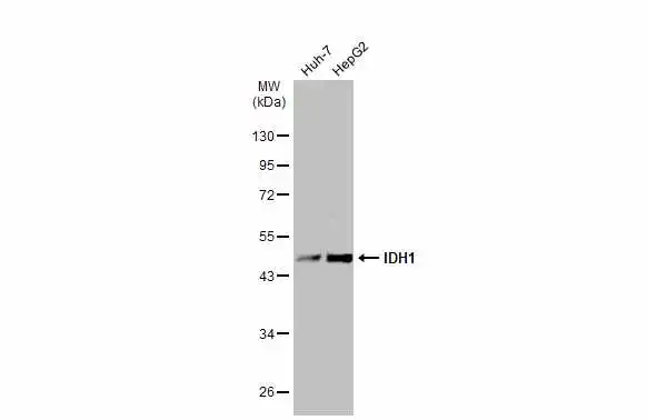
Various whole cell extracts (30 μg) were separated by 10% SDS-PAGE, and the membrane was blotted with IDH1 antibody (GTX105179) diluted at 1:1000. The HRP-conjugated anti-rabbit IgG antibody (GTX213110-01) was used to detect the primary antibody.

Wild-type (WT) and IDH1 knockout (KO) HeLa cell extracts (30 μg) were separated by 10% SDS-PAGE, and the membrane was blotted with IDH1 antibody (GTX105179) diluted at 1:500. The HRP-conjugated anti-rabbit IgG antibody (GTX213110-01) was used to detect the primary antibody, and the signal was developed with Trident ECL plus-Enhanced.

Various tissue extracts (50 μg) were separated by 10% SDS-PAGE, and the membrane was blotted with IDH1 antibody (GTX105179) diluted at 1:1000. The HRP-conjugated anti-rabbit IgG antibody (GTX213110-01) was used to detect the primary antibody.
IDH1 antibody detects IDH1 protein at cytoplasm by immunohistochemical analysis.Sample: Paraffin-embedded mouse brain.IDH1 stained by IDH1 antibody (GTX105179) diluted at 1:1000.Antigen Retrieval: Citrate buffer, pH 6.0, 15 min
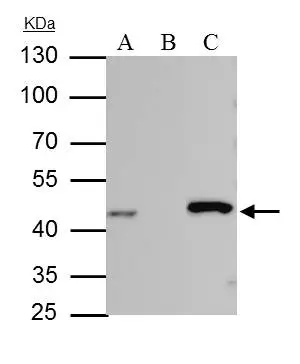
IDH-1 antibody immunoprecipitates IDH-1 protein in IP experiments. IP Sample: HepG2 whole cell lysate/extract A : 30 μg whole cell lysate/extract of IDH1 protein expressing HepG2 cells B : Control with 2.5 μg of pre-immune rabbit IgG C : Immunoprecipitation of IDH-1 protein by 2.5 μg of IDH-1 antibody (GTX105179) 10% SDS-PAGE The immunoprecipitated IDH-1 protein was detected by IDH-1 antibody (GTX105179) diluted at 1 : 1000. EasyBlot anti-rabbit IgG (HRP) (GTX221666-01) was used as a secondary reagent.
IDH1 antibody detects IDH1 protein at cytoplasm by immunohistochemical analysis.Sample: Paraffin-embedded rat colon.IDH1 stained by IDH1 antibody (GTX105179) diluted at 1:1000.Antigen Retrieval: Citrate buffer, pH 6.0, 15 min
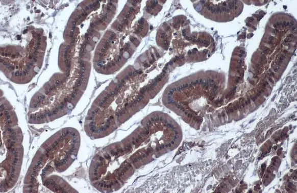
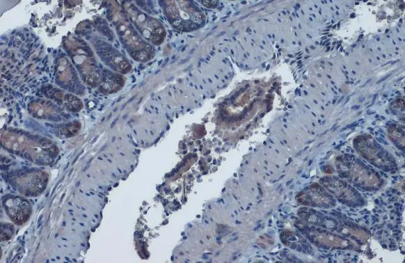
IDH1 antibody detects IDH1 protein at cytoplasm by immunohistochemical analysis.Sample: Paraffin-embedded rat duodenum.IDH1 stained by IDH1 antibody (GTX105179) diluted at 1:1000.Antigen Retrieval: Citrate buffer, pH 6.0, 15 min
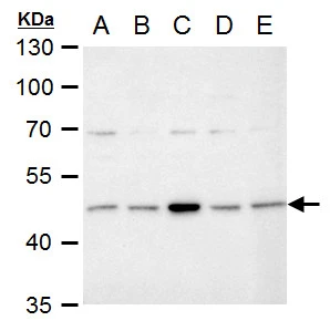
IDH1 antibody detects IDH1 protein by Western blot analysis.
A. 30 μg Neuro2A whole cell extract
B. 30 μg C8D30 whole cell extract
C. 30 μg NIH-3T3 whole cell extract
D. 30 μg Raw 264.7 whole cell extract
E. 30 μg C2Cl2 whole cell extract
10 % SDS-PAGE
IDH1 antibody (GTX105179) dilution: 1:1000
Immunohistochemical analysis of paraffin-embedded human colon carcinoma, using IDH1(GTX105179) antibody at 1:100 dilution.
Antigen Retrieval: Trilogy™ (EDTA based, pH 8.0) buffer, 15min
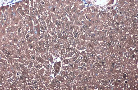
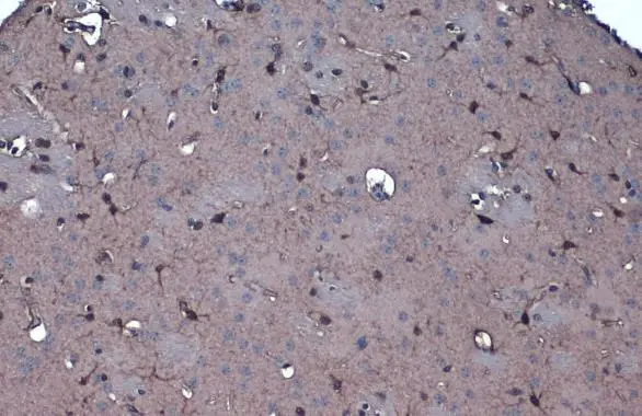
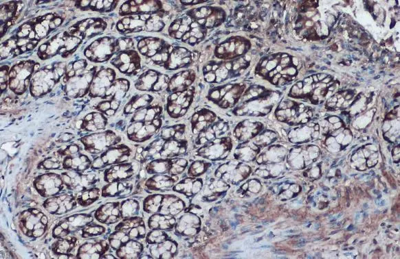
IDH1 antibody detects IDH1 protein at cytoplasm by immunohistochemical analysis.Sample: Paraffin-embedded mouse liver.IDH1 stained by IDH1 antibody (GTX105179) diluted at 1:1000.Antigen Retrieval: Citrate buffer, pH 6.0, 15 min
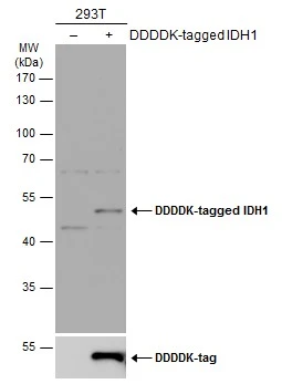
Non-transfected (–) and transfected (+) 293T whole cell extracts (30 μg) were separated by 10% SDS-PAGE, and the membrane was blotted with IDH1 antibody (GTX105179) diluted at 1:5000. The HRP-conjugated anti-rabbit IgG antibody (GTX213110-01) was used to detect the primary antibody.
IDH1 antibody detects IDH1 protein at cytoplasm by immunohistochemical analysis.Sample: Paraffin-embedded mouse intestine.IDH1 stained by IDH1 antibody (GTX105179) diluted at 1:1000.Antigen Retrieval: Citrate buffer, pH 6.0, 15 min
-
HostRabbit
-
ClonalityPolyclonal
-
IsotypeIgG
-
ApplicationsWB IHC-P IP
-
ReactivityHuman, Mouse, Rat