Integrin beta 1 / CD29 antibody


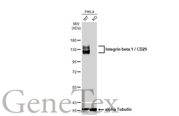
Wild-type (WT) and Integrin beta 1 / CD29 knockout (KO) HeLa cell extracts (30 μg) were separated by 7.5% SDS-PAGE, and the membrane was blotted with Integrin beta 1 / CD29 antibody (GTX128839) diluted at 1:400. The HRP-conjugated anti-rabbit IgG antibody (GTX213110-01) was used to detect the primary antibody.
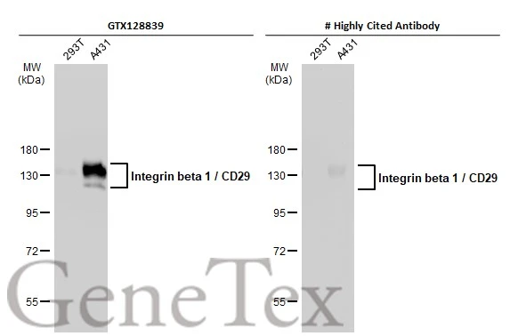
Various whole cell extracts (30 μg) were separated by 7.5% SDS-PAGE, and the membranes were blotted with Integrin beta 1 / CD29 antibody (GTX128839) diluted at 1:10000 and competitor's antibody diluted at 1:10000. The HRP-conjugated anti-rabbit IgG antibody (GTX213110-01) was used to detect the primary antibody.
*The competitor is not affiliated with GeneTex and does not endorse this product.
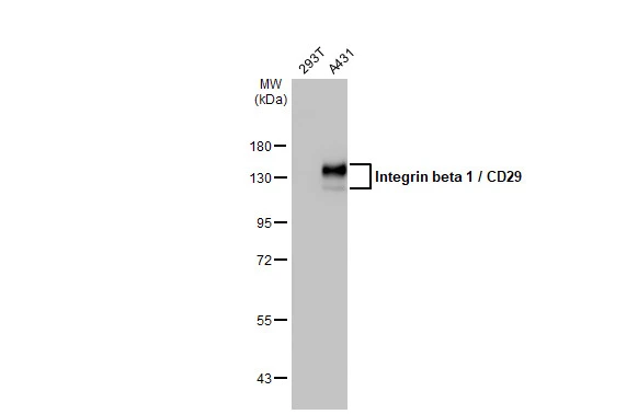
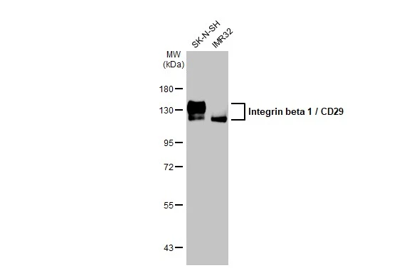
Various whole cell extracts (30 μg) were separated by 7.5% SDS-PAGE, and the membrane was blotted with Integrin beta 1 / CD29 antibody (GTX128839) diluted at 1:10000. The HRP-conjugated anti-rabbit IgG antibody (GTX213110-01) was used to detect the primary antibody.
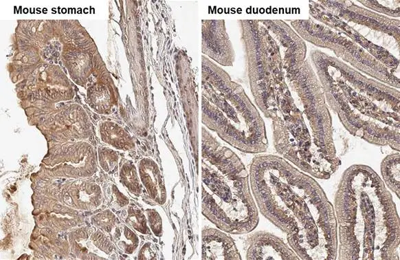
Integrin beta 1 / CD29 antibody detects Integrin beta 1 / CD29 protein by immunohistochemical analysis.Sample: Paraffin-embedded mouse tissues.Integrin beta 1 / CD29 stained by Integrin beta 1 / CD29 antibody (GTX128839) diluted at 1:500.Antigen Retrieval: Citrate buffer, pH 6.0, 15 min
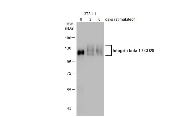
Unstimulatd and stimulatd 3T3-L1 whole cell extracts (20 μg) were separated by 7.5% SDS-PAGE, and the membrane was blotted with Integrin beta 1 / CD29 antibody (GTX128839) diluted at 1:500. The HRP-conjugated anti-rabbit IgG antibody (GTX213110-01) was used to detect the primary antibody. (The differentiation stimulated medium is composed by basal medium, 10% FBS, 50 ug/ml gentamicin, 1 nM L-glutamin, 500 uM IBMX, 1 uM dexamethasone, 2 uM rosiglitazone and 1 ug/ml insulin.)
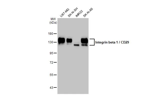
Various whole cell extracts (30 μg) were separated by 7.5% SDS-PAGE, and the membrane was blotted with Integrin beta 1 / CD29 antibody (GTX128839) diluted at 1:10000. The HRP-conjugated anti-rabbit IgG antibody (GTX213110-01) was used to detect the primary antibody.
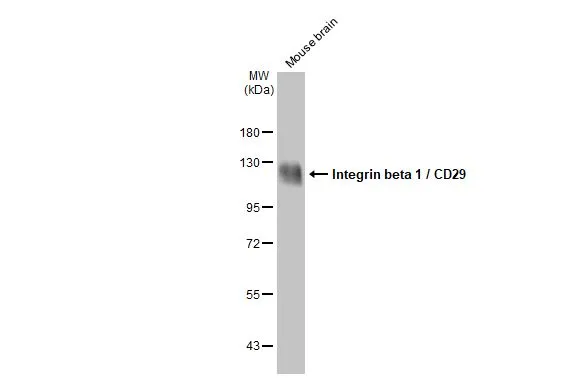
Mouse tissue extract (50 μg) was separated by 7.5% SDS-PAGE, and the membrane was blotted with Integrin beta 1 / CD29 antibody (GTX128839) diluted at 1:10000. The HRP-conjugated anti-rabbit IgG antibody (GTX213110-01) was used to detect the primary antibody.
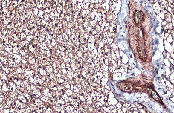
Integrin beta 1 / CD29 antibody detects Integrin beta 1 / CD29 protein at cytoplasm by immunohistochemical analysis.Sample: Paraffin-embedded mouse brown adipocyte.Integrin beta 1 / CD29 stained by Integrin beta 1 / CD29 antibody (GTX128839) diluted at 1:500.Antigen Retrieval: Citrate buffer, pH 6.0, 15 min
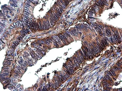
Integrin beta 1 / CD29 antibody detects Integrin beta 1 / CD29 protein at cell membrane and cytoplasm by immunohistochemical analysis.Sample: Paraffin-embedded human colon cancer.Integrin beta 1 / CD29 stained by Integrin beta 1 / CD29 antibody (GTX128839) diluted at 1:500.Antigen Retrieval: Citrate buffer, pH 6.0, 15 min
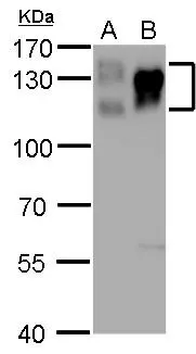
Integrin beta 1 / CD29 antibody detects Integrin beta 1 / CD29 protein by western blot analysis.
A. 30 μg PC-12 whole cell lysate/extract
B. 30 μg Rat-2 whole cell lysate/extract
7.5% SDS-PAGE
Integrin beta 1 / CD29 antibody (GTX128839) dilution: 1:5000
The HRP-conjugated anti-rabbit IgG antibody (GTX213110-01) was used to detect the primary antibody.
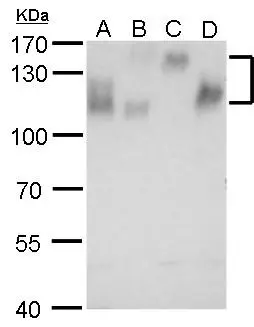
Integrin beta 1 / CD29 antibody detects Integrin beta 1 / CD29 protein by western blot analysis.
A. 30 μg NIH-3T3 whole cell lysate/extract
B. 30 μg BCL-1 whole cell lysate/extract
C. 30 μg Raw264.7 whole cell lysate/extract
D. 30 μg C2C12 whole cell lysate/extract
7.5% SDS-PAGE
Integrin beta 1 / CD29 antibody (GTX128839) dilution: 1:5000
The HRP-conjugated anti-rabbit IgG antibody (GTX213110-01) was used to detect the primary antibody.
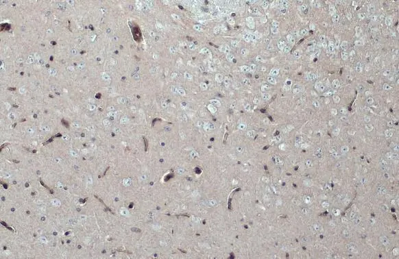
Integrin beta 1 / CD29 antibody detects Integrin beta 1 / CD29 protein at cytoplasm by immunohistochemical analysis.Sample: Paraffin-embedded mouse brain.Integrin beta 1 / CD29 stained by Integrin beta 1 / CD29 antibody (GTX128839) diluted at 1:500.Antigen Retrieval: Citrate buffer, pH 6.0, 15 min
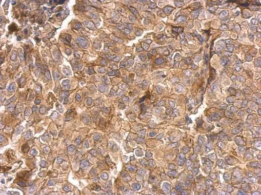
Integrin beta 1 / CD29 antibody detects Integrin beta 1 / CD29 protein on breast cancer by immunohistochemical analysis.
Sample: Paraffin-embedded breast cancer.
Integrin beta 1 / CD29 antibody (GTX128839) dilution: 1:500.
Antigen Retrieval: Trilogy™ (EDTA based, pH 8.0) buffer, 15min
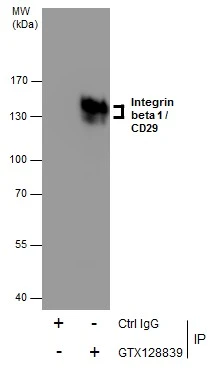
Immunoprecipitation of Integrin beta 1 / CD29 protein from HeLa whole cell extracts using 5 μg of Integrin beta 1 / CD29 antibody (GTX128839).
Western blot analysis was performed using Integrin beta 1 / CD29 antibody (GTX128839).
EasyBlot anti-Rabbit IgG (GTX221666-01) was used as a secondary reagent.
Various whole cell extracts (30 μg) were separated by 7.5% SDS-PAGE, and the membrane was blotted with Integrin beta 1 / CD29 antibody (GTX128839) diluted at 1:10000. The HRP-conjugated anti-rabbit IgG antibody (GTX213110-01) was used to detect the primary antibody.
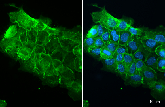
Integrin beta 1 / CD29 antibody detects Integrin beta 1 / CD29 protein at cell membrane by immunofluorescent analysis.Sample: A431 cells were fixed in ice-cold MeOH for 5 min.Green: Integrin beta 1 / CD29 stained by Integrin beta 1 / CD29 antibody (GTX128839) diluted at 1:500.Blue: Fluoroshield with DAPI (GTX30920).
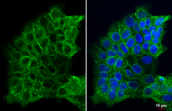
Integrin beta 1 / CD29 antibody detects Integrin beta 1 / CD29 protein at cell membrane by immunofluorescent analysis.Sample: A431 cells were fixed in ice-cold MeOH for 5 min.Green: Integrin beta 1 / CD29 stained by Integrin beta 1 / CD29 antibody (GTX128839) diluted at 1:500.Blue: Fluoroshield with DAPI (GTX30920).
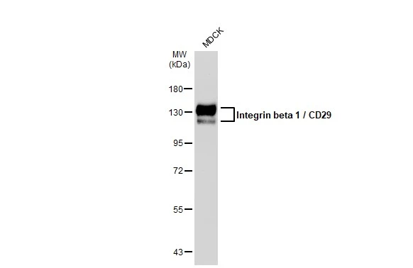
Whole cell extract (30 μg) was separated by 7.5% SDS-PAGE, and the membrane was blotted with Integrin beta 1 / CD29 antibody (GTX128839) diluted at 1:5000. The HRP-conjugated anti-rabbit IgG antibody (GTX213110-01) was used to detect the primary antibody.
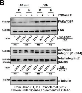
The data was published in the journal Oncotarget in 2017. PMID: 29050309
-
HostRabbit
-
ClonalityPolyclonal
-
IsotypeIgG
-
ApplicationsWB ICC/IF IHC-P IHC-Fr IP
-
ReactivityHuman, Mouse, Rat, Dog