Kv1.5 antibody
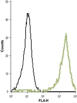
FACS analysis of THP-1 cells using GTX16716 Kv1.5 antibody.
Black : Unstained cell
Green : Cell staining with primary antibody
Dilution : 1:20
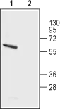
WB analysis of rat brain membrane lysate using GTX16716 Kv1.5 antibody preincubated with or without immunogen peptide.
Dilution : 1:200
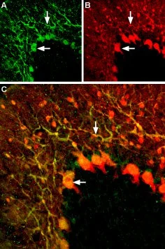
IHC-Fr analysis of rat cerebellum tissue using GTX16716 Kv1.5 antibody.
Panel A : KV1.5 (green) appears in both the soma of Purkinje cells (horizontal arrows) and in Purkinje dendrites (vertical arrows).
Panel B : Neurons expressing gamma amino butyric acid (GABA) were labeled with mouse anti-parvalbumin antibody (red).
Panel C : Merge of the two images demonstrates partial colocalization (white arrows).
Dilution : 1:200
-
HostRabbit
-
ClonalityPolyclonal
-
IsotypeIgG
-
ApplicationsWB IHC-Fr FCM
-
ReactivityHuman, Rat, Guinea pig