Lamin A + C antibody


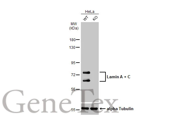
Wild-type (WT) and Lamin A + C knockout (KO) HeLa cell extracts (30 μg) were separated by 7.5% SDS-PAGE, and the membrane was blotted with Lamin A + C antibody (GTX101127) diluted at 1:1000. The HRP-conjugated anti-rabbit IgG antibody (GTX213110-01) was used to detect the primary antibody.
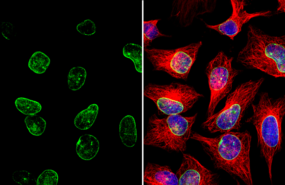
Lamin A + C antibody detects Lamin A + C protein at nuclear envelope by immunofluorescent analysis.Sample: HeLa cells were fixed in ice-cold MeOH for 5 min.Green: Lamin A + C stained by Lamin A + C antibody (GTX101127) diluted at 1:1000.Red: alpha Tubulin, stained by alpha Tubulin antibody [GT114] (GTX628802) diluted at 1:500.Blue: Fluoroshield with DAPI (GTX30920).
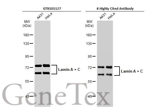
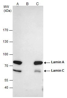
Various whole cell extracts (30 μg) were separated by 7.5% SDS-PAGE, and the membranes were blotted with Lamin A + C antibody (GTX101127) diluted at 1:500 and competitor's antibody diluted at 1:500. The HRP-conjugated anti-rabbit IgG antibody (GTX213110-01) was used to detect the primary antibody.
*The competitor is not affiliated with GeneTex and does not endorse this product.
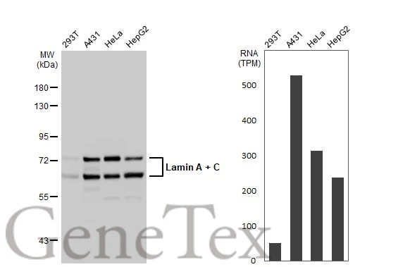
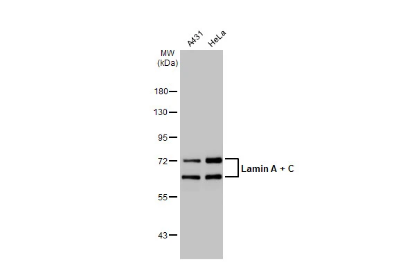
Various whole cell extracts (30 μg) were separated by 7.5% SDS-PAGE, and the membrane was blotted with Lamin A + C antibody (GTX101127) diluted at 1:2000. The HRP-conjugated anti-rabbit IgG antibody (GTX213110-01) was used to detect the primary antibody. Corresponding RNA expression data for the same cell lines are based on Human Protein Atlas program.
Various whole cell extracts (30 μg) were separated by 7.5% SDS-PAGE, and the membrane was blotted with Lamin A + C antibody (GTX101127) diluted at 1:5000. The HRP-conjugated anti-rabbit IgG antibody (GTX213110-01) was used to detect the primary antibody.
Immunohistochemical analysis of paraffin-embedded Mahlarvu xenograft, using Lamin A + C(GTX101127) antibody at 1:500 dilution.
Antigen Retrieval: Trilogy™ (EDTA based, pH 8.0) buffer, 15min
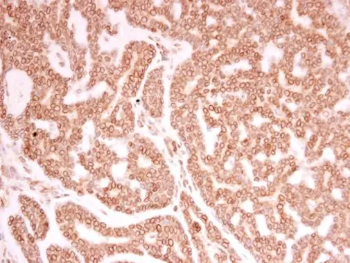
Lamin A + C antibody detects Lamin A + C protein at nuclear envelope in human breast carcinoma by immunohistochemical analysis.
Sample: Paraffin-embedded human breast carcinoma.
Lamin A + C antibody (GTX101127) diluted at 1:250.
Antigen Retrieval: Trilogy™ (EDTA based, pH 8.0) buffer, 15min
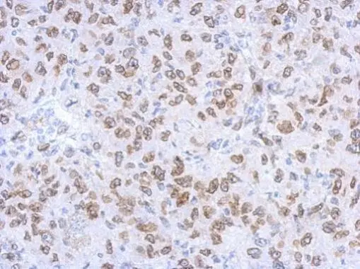
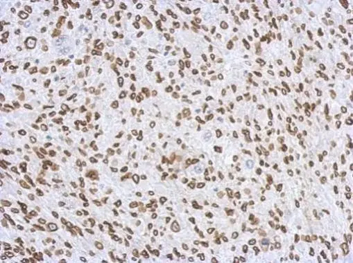
Immunohistochemical analysis of paraffin-embedded RT2 xenograft, using Lamin A + C(GTX101127) antibody at 1:500 dilution.
Antigen Retrieval: Trilogy™ (EDTA based, pH 8.0) buffer, 15min
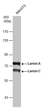
Sample (30 μg of whole cell lysate)
A: NIH-3T3
7.5% SDS PAGE
GTX101127 diluted at 1:1000
The HRP-conjugated anti-rabbit IgG antibody (GTX213110-01) was used to detect the primary antibody.
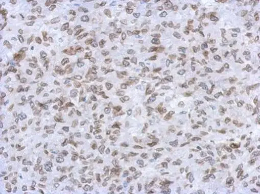
Immunohistochemical analysis of paraffin-embedded C2C12 xenograft, using Lamin A + C(GTX101127) antibody at 1:500 dilution.
Antigen Retrieval: Trilogy™ (EDTA based, pH 8.0) buffer, 15min
Lamin A + C antibody immunoprecipitates Lamin A + C protein in IP experiments.
IP samples: HeLa whole cell extract
A. 50 μg HeLa whole cell extract
B. Control with 4 μg of preimmune Rabbit IgG
C. Immunoprecipitation of Lamin A + C protein by 4 μg Lamin A + C antibody (GTX101127)
7.5 % SDS-PAGE
The immunoprecipitated Lamin A + C protein was detected by Lamin A + C antibody (GTX101127) diluted at 1:500.
[EasyBlot anti-rabbit IgG (GTX221666-01) was used as a secondary reagent]
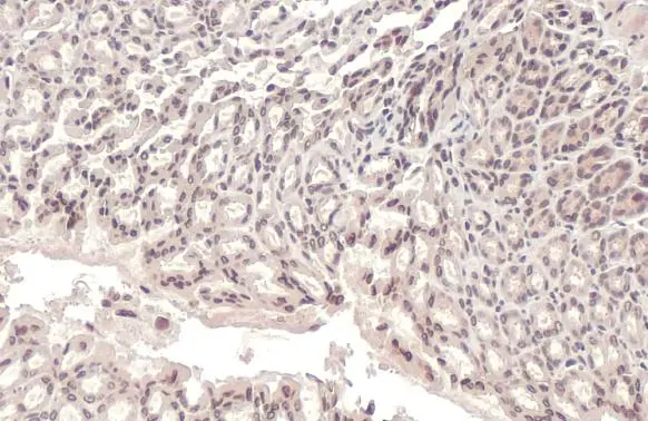
Lamin A + C antibody detects Lamin A + C protein at cytoplasm and nucleus by immunohistochemical analysis.
Sample: Paraffin-embedded mouse stomach.
Lamin A + C stained by Lamin A + C antibody (GTX101127) diluted at 1:100.
Antigen Retrieval: Citrate buffer, pH 6.0, 15 min
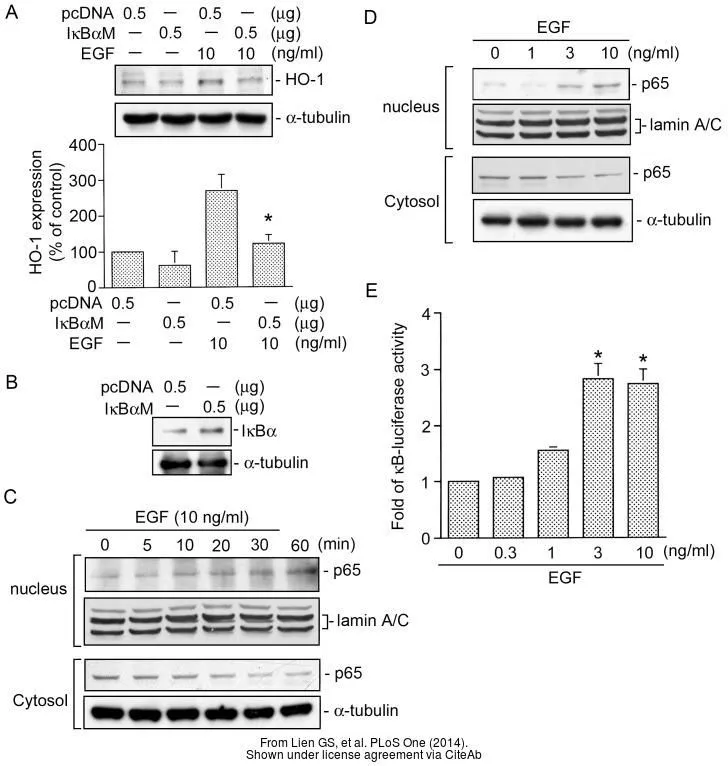
The data was published in the journal PLoS One in 2014. PMID: 25122478
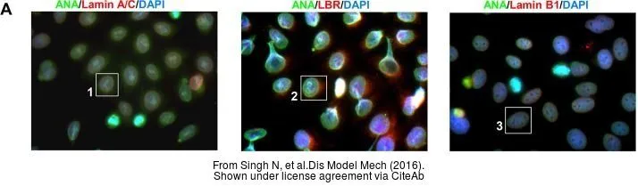
The data was published in the journal Dis Model Mech in 2016. PMID: 27483354
-
HostRabbit
-
ClonalityPolyclonal
-
IsotypeIgG
-
ApplicationsWB ICC/IF IHC-P IP
-
ReactivityHuman, Mouse, Rat