MAP2 antibody

Mouse tissue extract (50 μg) was separated by 5% SDS-PAGE, and the membrane was blotted with MAP2 antibody (GTX133109) diluted at 1:50000. The HRP-conjugated anti-rabbit IgG antibody (GTX213110-01) was used to detect the primary antibody.
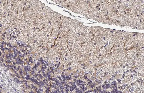
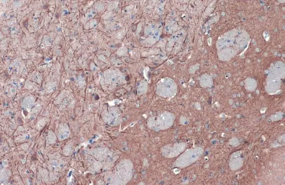
MAP2 antibody detects MAP2 protein at cytoplasm by immunohistochemical analysis.Sample: Paraffin-embedded mouse cerebellum.MAP2 stained by MAP2 antibody (GTX133109) diluted at 1:500.Antigen Retrieval: Citrate buffer, pH 6.0, 15 min
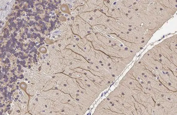
MAP2 antibody detects MAP2 protein at cytoplasm by immunohistochemical analysis.Sample: Paraffin-embedded rat cerebellum.MAP2 stained by MAP2 antibody (GTX133109) diluted at 1:500.Antigen Retrieval: Citrate buffer, pH 6.0, 15 min
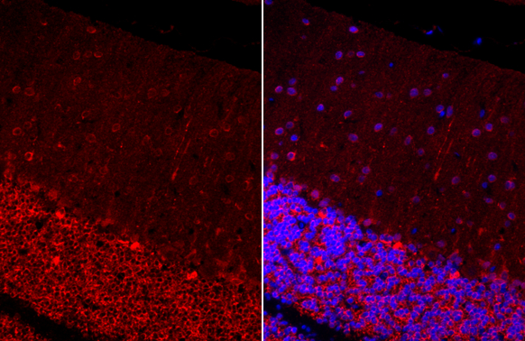
MAP2 antibody detects MAP2 protein at cytoplasm by immunohistochemical analysis.Sample: Paraffin-embedded rat cerebellum.Green: MAP2 stained by MAP2 antibody (GTX133109) diluted at 1:250.Blue: Fluoroshield with DAPI (GTX30920).Antigen Retrieval: Citrate buffer, pH 6.0, 15 min
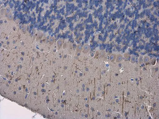
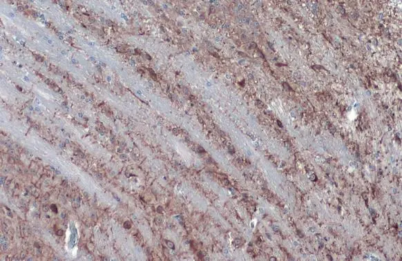
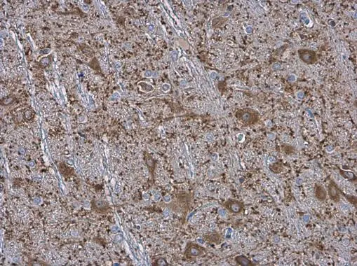
MAP2 antibody detects MAP2 protein at cytoplasm in mouse brain by immunohistochemical analysis.
Sample: Paraffin-embedded mouse brain.
MAP2 antibody (GTX133109) diluted at 1:500.
Antigen Retrieval: Citrate buffer, pH 6.0, 15 min
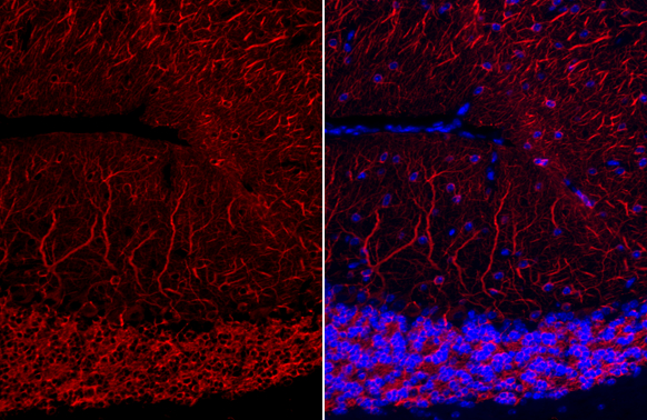
MAP2 antibody detects MAP2 protein at cytoplasm by immunohistochemical analysis.Sample: Paraffin-embedded mouse cerebellum.Green: MAP2 stained by MAP2 antibody (GTX133109) diluted at 1:250.Blue: Fluoroshield with DAPI (GTX30920).Antigen Retrieval: Citrate buffer, pH 6.0, 15 min
MAP2 antibody detects MAP2 protein at cytoplasm by immunohistochemical analysis.Sample: Paraffin-embedded mouse brain.MAP2 stained by MAP2 antibody (GTX133109) diluted at 1:2000.Antigen Retrieval: Citrate buffer, pH 6.0, 15 min
MAP2 antibody detects MAP2 protein at cytoplasm by immunohistochemical analysis.Sample: Paraffin-embedded mouse brain.MAP2 stained by MAP2 antibody (GTX133109) diluted at 1:2000.Antigen Retrieval: Citrate buffer, pH 6.0, 15 min
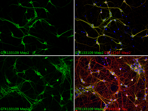
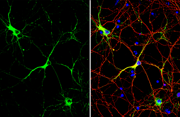
MAP2 antibody detects MAP2 protein in dendrites, but not in axons, by immunofluorescent analysis .
Sample: DIV9 rat E18 primary cortical neurons were fixed in 4% paraformaldehyde at RT for 15 min.
Grenn: MAP2 protein stained by MAP2 antibody (GTX133109) diluted at 1:500.
Red (Top right): Dendrites, stained by MAP2 antibody [HM-2] (GTX11267) diluted at 1:1000.
Red (Bottom right): Axons, stained by Tau antibody (GTX49353 ) diluted at 1:500.
Blue: Fluoroshield with DAPI (GTX30920).
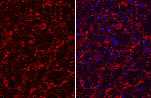
MAP2 antibody detects MAP2 protein at cytoplasm in rat brain by immunohistochemical analysis.
Sample: Paraffin-embedded rat brain.
MAP2 antibody (GTX133109) diluted at 1:500.
Antigen Retrieval: Citrate buffer, pH 6.0, 15 min
MAP2 antibody detects MAP2 protein in dendrites, but not in axons, by immunofluorescent analysis. Sample: DIV10 rat E18 primary cortical neuron cells were fixed in 4% paraformaldehyde at RT for 15 min.Green: MAP2 stained by MAP2 antibody (GTX133109) diluted at 1:500.Red: Tau, stained by Tau antibody [GT287] (GTX634809) diluted at 1:500.Blue: Fluoroshield with DAPI (GTX30920).
MAP2 antibody detects MAP2 protein at cytoplasm by immunohistochemical analysis.Sample: Paraffin-embedded rat cerebellum.Green: MAP2 stained by MAP2 antibody (GTX133109) diluted at 1:250.Blue: Fluoroshield with DAPI (GTX30920).Antigen Retrieval: Citrate buffer, pH 6.0, 15 min
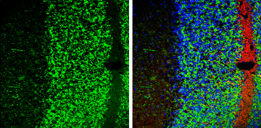
MAP2 antibody detects MAP2 protein expression by immunohistochemical analysis.
Sample: Frozen-sectioned adult mouse cerebellum.
Green: MAP2 protein stained by MAP2 antibody (GTX133109) diluted at 1:250.
Red: Tau, stained by Tau antibody (GTX49353) diluted at 1:500.
Blue: Fluoroshield with DAPI (GTX30920).
-
HostRabbit
-
ClonalityPolyclonal
-
IsotypeIgG
-
ApplicationsWB ICC/IF IHC-P IHC-Fr
-
ReactivityHuman, Mouse, Rat, Fish