MEK1 antibody


MEK1 antibody detects MEK1 protein at cytoplasm by immunohistochemical analysis.Sample: Paraffin-embedded mouse liver.MEK1 stained by MEK1 antibody (GTX134234) diluted at 1:1000.Antigen Retrieval: Citrate buffer, pH 6.0, 15 min
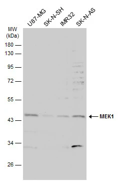
Various whole cell extracts (30 μg) were separated by 10% SDS-PAGE, and the membrane was blotted with MEK1 antibody (GTX134234) diluted at 1:1000. The HRP-conjugated anti-rabbit IgG antibody (GTX213110-01) was used to detect the primary antibody.
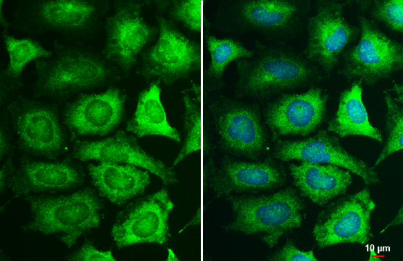
MEK1 antibody detects MEK1 protein at cytoplasm by immunofluorescent analysis.Sample: HeLa cells were fixed in 4% paraformaldehyde at RT for 15 min.Green: MEK1 stained by MEK1 antibody (GTX134234) diluted at 1:500.Blue: Hoechst 33342 staining.Scale bar= 10 μm.
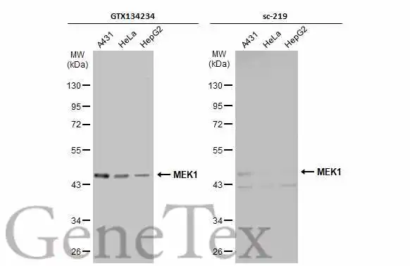
Various whole cell extracts (30 μg) were separated by 10% SDS-PAGE, and the membranes were blotted with MEK1 antibody (GTX134234) diluted at 1:500 and competitor's antibody (sc-219) diluted at 1:500. The HRP-conjugated anti-rabbit IgG antibody (GTX213110-01) was used to detect the primary antibody, and the signal was developed with Trident ECL plus-Enhanced.
*The competitor is not affiliated with GeneTex and does not endorse this product.
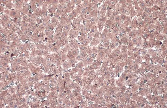
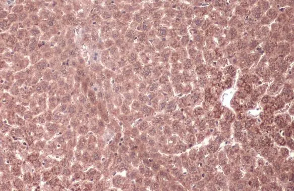
MEK1 antibody detects MEK1 protein at cytoplasm by immunohistochemical analysis.Sample: Paraffin-embedded rat liver.MEK1 stained by MEK1 antibody (GTX134234) diluted at 1:1000.Antigen Retrieval: Citrate buffer, pH 6.0, 15 min
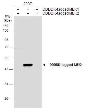
Non-transfected (–) and transfected (+) 293T whole cell extracts (30 μg) were separated by 10% SDS-PAGE, and the membrane was blotted with MEK1 antibody (GTX134234) diluted at 1:5000. The HRP-conjugated anti-rabbit IgG antibody (GTX213110-01) was used to detect the primary antibody.
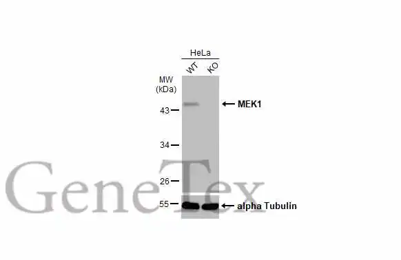
Wild-type (WT) and MEK1 knockout (KO) HeLa cell extracts (30 μg) were separated by 10% SDS-PAGE, and the membrane was blotted with MEK1 antibody (GTX134234) diluted at 1:1000. The HRP-conjugated anti-rabbit IgG antibody (GTX213110-01) was used to detect the primary antibody, and the signal was developed with Trident ECL plus-Enhanced.
-
HostRabbit
-
ClonalityPolyclonal
-
IsotypeIgG
-
ApplicationsWB ICC/IF IHC-P IHC-Fr
-
ReactivityHuman, Mouse, Rat