Oct4 antibody


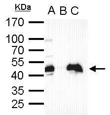
Oct4 antibody immunoprecipitates Oct4 protein in IP experiments. IP Sample: cell lysate/extract of Oct4 gene transfected 293T cells A. Cell lysate/extract of transfected 293T cell B. Control with 2 μg of preimmune rabbit IgG C. Immunoprecipitation of Oct4 by 2 μg of Oct4 antibody (GTX101497) 12% SDS-PAGE The immunoprecipitated Oct4 protein was detected by Oct4 antibody (GTX101497) diluted at 1:1000. EasyBlot anti-rabbit IgG (GTX221666-01) was used as a secondary reagent.
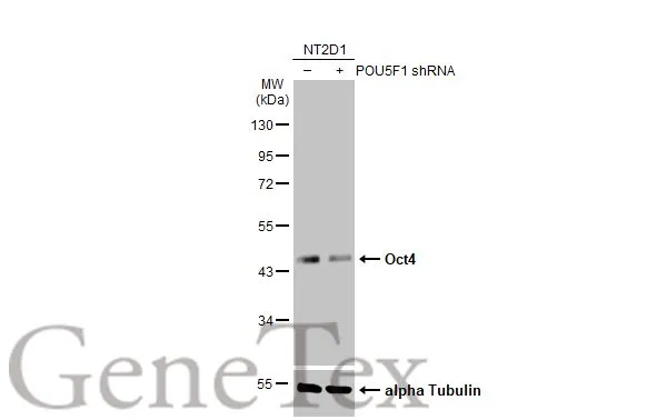
Non-transfected (–) and transfected (+) NT2D1 whole cell extracts (30 μg) were separated by 10% SDS-PAGE, and the membrane was blotted with Oct4 antibody (GTX101497) diluted at 1:200000. The HRP-conjugated anti-rabbit IgG antibody (GTX213110-01) was used to detect the primary antibody.
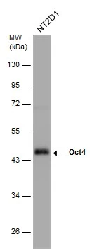
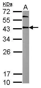
Whole cell extract (30 μg) was separated by 10% SDS-PAGE, and the membrane was blotted with Oct4 antibody (GTX101497) diluted at 1:100000.
Oct4 antibody detects Oct4 protein by western blot analysis.
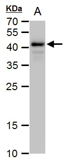
A. 30 μg human ESC whole cell extract
12% SDS-PAGE
Oct4 antibody (GTX101497) dilution: 1:10000
The HRP-conjugated anti-rabbit IgG antibody (GTX213110-01) was used to detect the primary antibody.
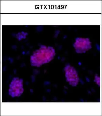
Immunofluorescence analysis of mESCs on CF1 feeders, using OCT3/4(GTX101497) antibody at 1:200 dilution.
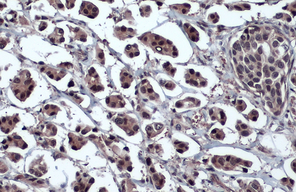
Oct4 antibody detects Oct4 protein at nucleus by immunohistochemical analysis.Sample: Paraffin-embedded human breast carcinoma.Oct4 stained by Oct4 antibody (GTX101497) diluted at 1:1000.Antigen Retrieval: Citrate buffer, pH 6.0, 15 min
Sample (20 μg of whole cell lysate)
A: mouse ESC
12% SDS PAGE
GTX101497 diluted at 1:10000
The HRP-conjugated anti-rabbit IgG antibody (GTX213110-01) was used to detect the primary antibody.
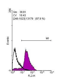
Flow cytometry on human embryonic stem cells, staining with Oct4 (GTX101497)antibody at 1:100 dilution(purple) or rabbit IgG (black).
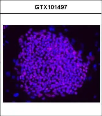
Immunofluorescence analysis of hESCs on CF1 feeders, using OCT3/4(GTX101497) antibody at 1:200 dilution.
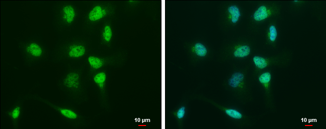
Oct4 antibody detects Oct4 protein at nucleus by immunofluorescent analysis.
Sample: NT2D1 cells were fixed in 4% paraformaldehyde at RT for 15 min.
Green: Oct4 protein stained by Oct4 antibody (GTX101497) diluted at 1:500.
Blue: Hoechst 33342 staining.
Scale bar = 10 μm.
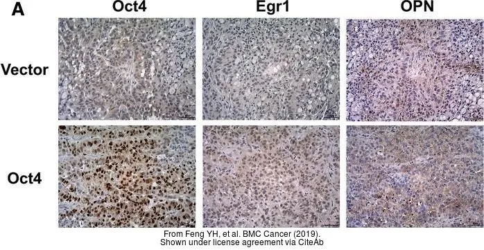
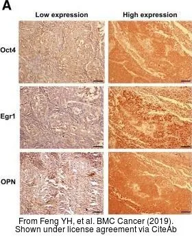
The data was published in the journal BMC Cancer in 2019. PMID: 31399076
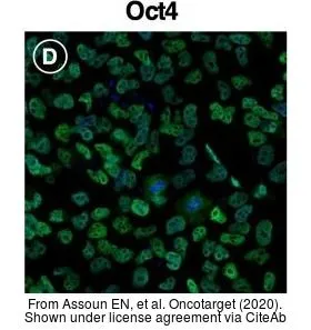
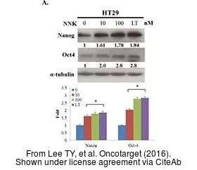
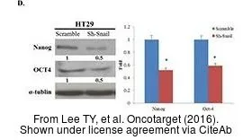
The data was published in the journal Oncotarget in 2020.PMID: 32256979
-
HostRabbit
-
ClonalityPolyclonal
-
IsotypeIgG
-
ApplicationsWB ICC/IF IHC-P IHC-Fr FCM IP
-
ReactivityHuman, Mouse, Rat