Orexin Receptor 1 antibody
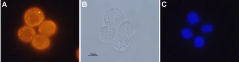
ICC/IF analysis of PFA-fixed human colon cancer cells using GTX54847 Orexin Receptor 1 antibody.
Panel A : Primary antibody
Panel B : Live view of the same field as in (A)
Panel C : DNA dye Hoechst 33342
Dilution : 1:200
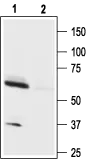
WB analysis of mouse brain tissue lysate using GTX54847 Orexin Receptor 1 antibody preincubated with or without immunogen peptide.
Dilution : 1:200
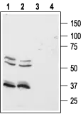
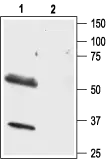
WB analysis of Colo-205 (lanes 1 and 3) and HT-29 (lanes 2 and 4) cell lysates using GTX54847 Orexin Receptor 1 antibody preincubated with or without immunogen peptide.
Dilution : 1:500
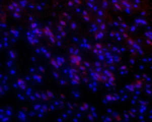
IHC-Fr analysis of rat brain tissue using GTX54847 Orexin Receptor 1 antibody.
Red : Primary antibody
Blue : Hoechst 33342 is used as a counterstain
Dilution : 1:500
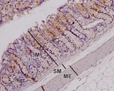
IHC-P analysis of rat colon tissue using GTX54847 Orexin Receptor 1 antibody. Note that the stain (red-brown color) is highly specific for absorptive cells in the superior third of the intestinal glands. Immunolabeling was detected using DAB as the chromogen and hematoxilin as the counterstain.
Dilution : 1:100
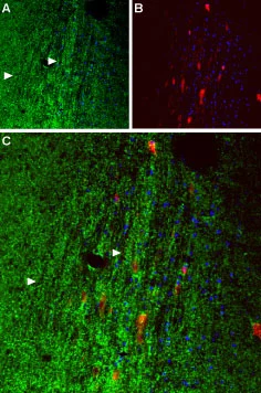
IHC-Fr analysis of mouse septum tissue using GTX54847 Orexin Receptor 1 antibody. Cell nuclei (blue) are visualized with Hoechst 33342.
Panel A : OX1R (green) appears in axonal processes (right-pointing triangles).
Panel B : Parvalbumin (red) appears in septal neurons.
Panel C : Merge of OX1R and parvalbumin suggests that orexinergic innervation covers the entire septal nucleus rather than restricted to individual neurons.
Dilution : 1:50
WB analysis of rat brain lysate using GTX54847 Orexin Receptor 1 antibody preincubated with or without immunogen peptide.
Dilution : 1:500
-
HostRabbit
-
ClonalityPolyclonal
-
IsotypeIgG
-
ApplicationsWB ICC/IF IHC-P IHC-Fr
-
ReactivityHuman, Mouse, Rat