P2Y12 antibody
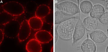
Live cell imaging analysis of intact living RBL cells using GTX16888 P2Y12 antibody.
Panel A : Primary antibody
Panel B : Live view of the cell
Dilution : 1:100
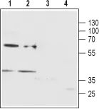
WB analysis of rat brain membrane (lanes 1 and 3) and MEG-01 (lanes 2 and 4) lysates using GTX16888 P2Y12 antibody preincubated with or without immunogen peptide.
Dilution : 1:200
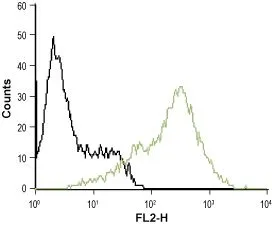
FACS analysis of MEG-O1 cells using GTX16888 P2Y12 antibody.
Black : Unstained cell
Green : Cell staining with primary antibody
Antibody amount : 5-10 μg antibody / 1 x 10⁶ cells
-
HostRabbit
-
ClonalityPolyclonal
-
IsotypeIgG
-
ApplicationsWB ICC/IF FCM LCI
-
ReactivityHuman, Rat