PAX6 antibody


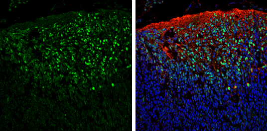
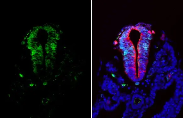
PAX6 antibody detects PAX6 protein expression by immunohistochemical analysis.
Sample: Frozen sectioned E13.5 Rat brain.
Green: PAX6 protein stained by PAX6 antibody (GTX113241) diluted at 1:250.
Red: beta Tubulin 3/ TUJ1, a mature neuron marker, stained by beta Tubulin 3/ TUJ1 antibody [GT11710] (GTX631836) diluted at 1:500.
Blue: Fluoroshield with DAPI (GTX30920).
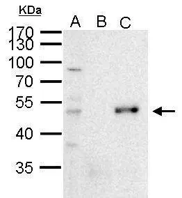
PAX6 antibody immunoprecipitates PAX6 protein in IP experiments. IP Sample: 1000 μg 293T whole cell lysate/extract A. 50 μg 293T whole cell lysate/extract B. Control with 2 μg of preimmune rabbit IgG C. Immunoprecipitation of PAX6 protein by 2 μg of PAX6 antibody (GTX113241) 10% SDS-PAGE The immunoprecipitated PAX6 protein was detected by PAX6 antibody (GTX113241) diluted at 1:1000. EasyBlot anti-rabbit IgG (GTX221666-01) was used as a secondary reagent.
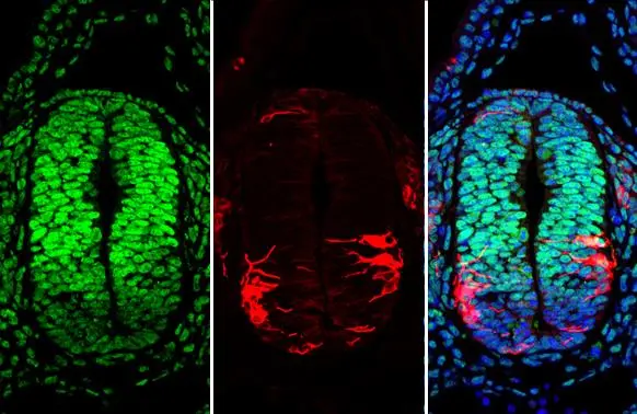
PAX6 antibody detects PAX6 protein at nucleus by immunohistochemical analysis.Sample: Paraffin-embedded mouse E10.5 embryo.Green: PAX6 stained by PAX6 antibody (GTX113241) diluted at 1:250.Red: beta Tubulin 3/ Tuj1, a cytoskeleton marker, stained by beta Tubulin 3/ Tuj1 antibody [GT11710] (GTX631836) diluted at 1:500.Blue: Fluoroshield with DAPI (GTX30920).Antigen Retrieval: Citrate buffer, pH 6.0, 15 min
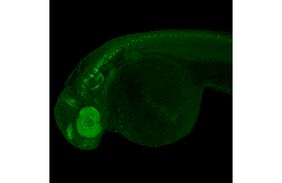
PAX6 antibody detects PAX6 protein on whole mount zebrafish by immunohistochemical analysis.Sample: Paraformaldehyde-fixed 2 days-post-fertilization zebrafish embryo.Green: PAX6 stained by PAX6 antibody (GTX113241) diluted at 1:300.Antigen Retrieval: Tris-HCl buffer, pH 9.0, 20 min at 70ºC
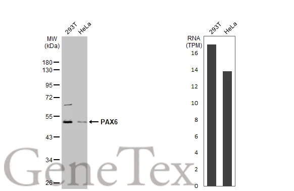
Various whole cell extracts (30 μg) were separated by 10% SDS-PAGE, and the membrane was blotted with PAX6 antibody (GTX113241) diluted at 1:1000. The HRP-conjugated anti-rabbit IgG antibody (GTX213110-01) was used to detect the primary antibody. Corresponding RNA expression data for the same cell lines are based on Human Protein Atlas program.
PAX6 antibody detects PAX6 protein by immunohistochemical analysis.Sample: Paraffin-embedded mouse E10.5 embryo.Green: PAX6 stained by PAX6 antibody (GTX113241) diluted at 1:500.Red: beta Tubulin 3/ Tuj1, a Cytoskeleton marker, stained by beta Tubulin 3/ Tuj1 antibody [GT11710] (GTX631836) diluted at 1:500.Blue: Hoechst 33342 staining.Antigen Retrieval: Citrate buffer, pH 6.0, 15 min
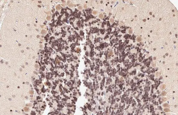
PAX6 antibody detects PAX6 protein at nucleus by immunohistochemical analysis.Sample: Paraffin-embedded rat cerebellum.PAX6 stained by PAX6 antibody (GTX113241) diluted at 1:500.Antigen Retrieval: Citrate buffer, pH 6.0, 15 min
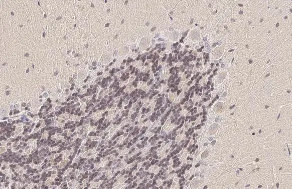
PAX6 antibody detects PAX6 protein at nucleus by immunohistochemical analysis.Sample: Paraffin-embedded mouse cerebellum.PAX6 stained by PAX6 antibody (GTX113241) diluted at 1:500.Antigen Retrieval: Citrate buffer, pH 6.0, 15 min
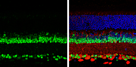
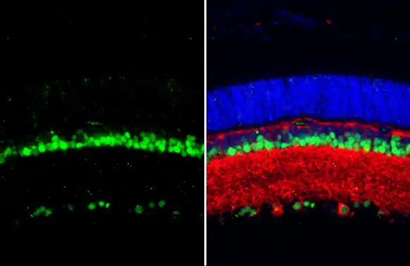
PAX6 antibody detects PAX6 protein by immunohistochemical analysis.
Samples: Paraffin-Embedded mouse retina.
Green: PAX6 protein stained by PAX6 antibody (GTX113241) diluted at 1:250.
Red: beta Tubulin 3/ Tuj1, stained by beta Tubulin 3/ Tuj1 antibody [GT1338] (GTX631831) diluted at 1:500.
Blue: Fluoroshield with DAPI (GTX30920).
Antigen Retrieval: Citrate buffer, pH 6.0, 15 min
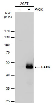
Non-transfected (–) and transfected (+) 293T whole cell extracts (30 μg) were separated by 10% SDS-PAGE, and the membrane was blotted with PAX6 antibody (GTX113241) diluted at 1:1000. The HRP-conjugated anti-rabbit IgG antibody (GTX213110-01) was used to detect the primary antibody.
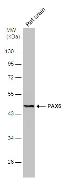
Rat tissue extract (50 μg) was separated by 10% SDS-PAGE, and the membrane was blotted with PAX6 antibody (GTX113241) diluted at 1:500. The HRP-conjugated anti-rabbit IgG antibody (GTX213110-01) was used to detect the primary antibody.
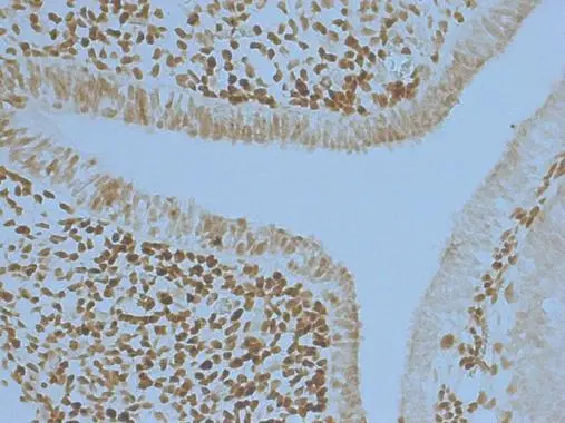
PAX6 antibody detects PAX6 protein at nucleus in embryonic mouse brain by immunohistochemical analysis.
Sample: Frozen section of embryonic mouse brain (mE12.5).
PAX6 antibody (GTX113241) diluted at 1:250.
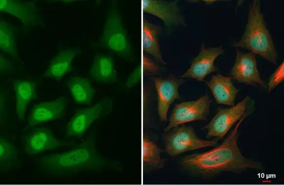
PAX6 antibody detects PAX6 protein at cytoplasm and nucleus by immunofluorescent analysis.Sample: HeLa cells were fixed in 4% paraformaldehyde at RT for 15 min.Green: PAX6 stained by PAX6 antibody (GTX113241) diluted at 1:500.Red: alpha Tubulin, a cytoskeleton marker, stained by alpha Tubulin antibody [GT114] (GTX628802) diluted at 1:1000.Blue: Fluoroshield with DAPI (GTX30920).
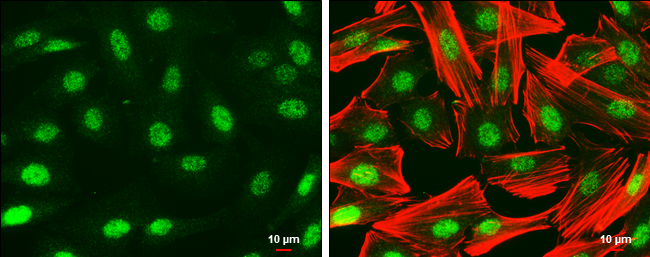
PAX6 antibody detects PAX6 protein at nucleus by immunofluorescent analysis.
Sample: SK-N-SH cells were fixed in 4% paraformaldehyde at RT for 15 min.
Green: PAX6 protein stained by PAX6 antibody (GTX113241) diluted at 1:500.
Red: Phalloidin, a cytoskeleton marker, diluted at 1:50.
Scale bar = 10 μm.
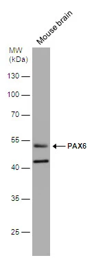
Mouse tissue extract (50 μg) was separated by 10% SDS-PAGE, and the membrane was blotted with PAX6 antibody (GTX113241) diluted at 1:1000. The HRP-conjugated anti-rabbit IgG antibody (GTX213110-01) was used to detect the primary antibody.
PAX6 antibody detects PAX6 protein at nucleus by immunohistochemical analysis.Sample: Paraffin-embedded rat eye.Green: PAX6 stained by PAX6 antibody (GTX113241) diluted at 1:250.Red: beta Tubulin 3/ Tuj1, a neural marker, stained by beta Tubulin 3/ Tuj1 antibody [GT11710] (GTX631836) diluted at 1:500.Blue: Fluoroshield with DAPI (GTX30920).Antigen Retrieval: Citrate buffer, pH 6.0, 15 min
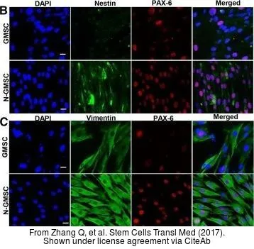
The data was published in the journal Stem Cells Transl Med in 2017. PMID: 28191764
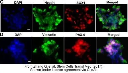
The data was published in the journal Stem Cells Transl Med in 2017. PMID: 28191764
-
HostRabbit
-
ClonalityPolyclonal
-
IsotypeIgG
-
ApplicationsWB ICC/IF IHC-P IHC-Fr IHC-Wm IP
-
ReactivityHuman, Mouse, Rat, Zebrafish