PEX14 antibody

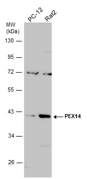
Various whole cell extracts (30 μg) were separated by 10% SDS-PAGE, and the membrane was blotted with PEX14 antibody (GTX129230) diluted at 1:1000. The HRP-conjugated anti-rabbit IgG antibody (GTX213110-01) was used to detect the primary antibody.
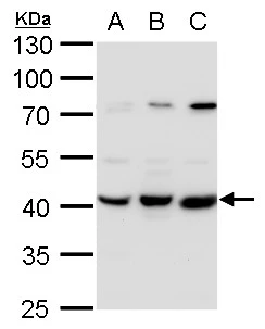
PEX14 antibody detects PEX14 protein by western blot analysis.
A. 30 μg 293T whole cell lysate/extract
B. 30 μg A431 whole cell lysate/extract
C. 30 μg HeLa whole cell lysate/extract
10% SDS-PAGE
PEX14 antibody (GTX129230) dilution: 1:1000
The HRP-conjugated anti-rabbit IgG antibody (GTX213110-01) was used to detect the primary antibody.
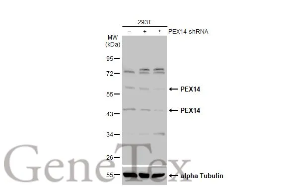
Non-transfected (–) and transfected (+) 293T whole cell extracts (30 μg) were separated by 10% SDS-PAGE, and the membrane was blotted with PEX14 antibody (GTX129230) diluted at 1:500. The HRP-conjugated anti-rabbit IgG antibody (GTX213110-01) was used to detect the primary antibody.
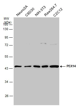
Various whole cell extracts (30 μg) were separated by 10% SDS-PAGE, and the membrane was blotted with PEX14 antibody (GTX129230) diluted at 1:1000. The HRP-conjugated anti-rabbit IgG antibody (GTX213110-01) was used to detect the primary antibody.
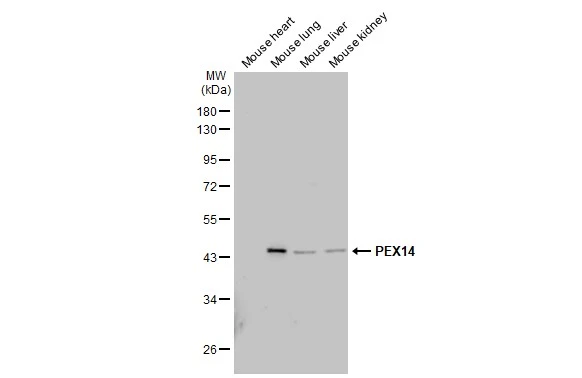
Various tissue extracts (50 μg) were separated by 10% SDS-PAGE, and the membrane was blotted with PEX14 antibody (GTX129230) diluted at 1:1000. The HRP-conjugated anti-rabbit IgG antibody (GTX213110-01) was used to detect the primary antibody.
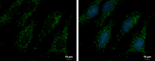
PEX14 antibody detects PEX14 protein at peroxisome by immunofluorescent analysis.
Sample: HeLa cells were fixed in 4% paraformaldehyde at RT for 15 min.
Green: PEX14 protein stained by PEX14 antibody (GTX129230) diluted at 1:500.
Blue: Hoechst 33342 staining.
-
HostRabbit
-
ClonalityPolyclonal
-
IsotypeIgG
-
ApplicationsWB ICC/IF
-
ReactivityHuman, Mouse, Rat