RNF2 antibody
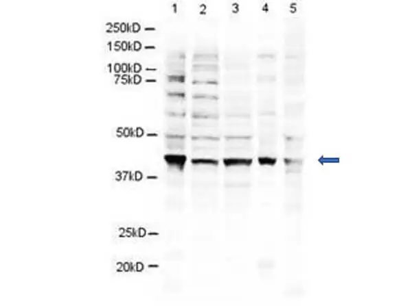
WB analysis of various samples using GTX48486 RNF2 antibody.
Lane 1 : NIH-3T3 whole cell lysate
Lane 2 : U937 whole cell lysate
Lane 3 : Jurkat whole cell lysate
Lane 4 : Mouse brain tissue lysate
Lane 5 : CHO-K1 whole cell lysate
Loading : 20 μg
Dilution : 1:500
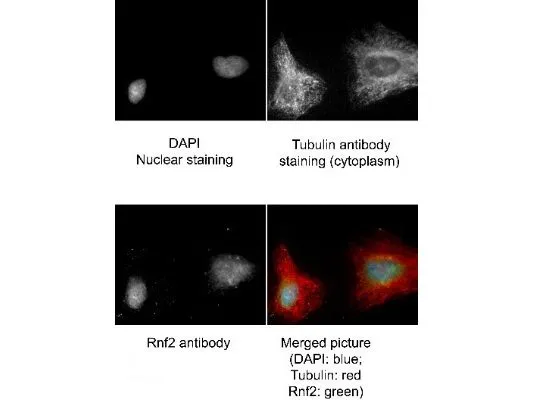
Immunofluorescence Microscopy of Goat anti-RING1B antibody (GTX48486). Tissue: human HeLa cells. Fixation: methanol and blocked with 0.2% fish scale gelatin for 1 hour at 25ºC. Antigen retrieval: not required. Primary antibody: RING1B antibody at 1:300 for 20 minutes at 25ºC. Secondary antibody: Alexa FluorR488-conjugated Donkey anti-goat IgG secondary antibody at 1:500 for 45 min at RT. Localization: RING1B is nuclear and occasionally cytoplasmic. Staining: RING1B (RNF2) as green signal, Tubulin cytoplasm staining red, and DAPI (blue) nuclear counterstain.
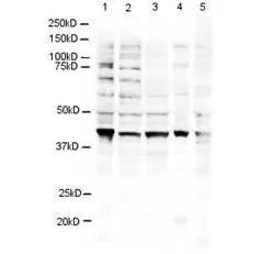
Western blot using GeneTex's Affinity Purified anti-RING1B antibody shows detection of a 38 kDa band corresponding to human RING1B in 3T3 (lane 1), U937 (lane 2), Jurkat (lane 3), mouse brain (lane 4) and CHO-K1 (lane 5) cell lysates. Approximately 20 μg of lysate was run on a SDS-PAGE and transferred onto nitrocellulose followed by reaction with a 1:500 dilution of anti-RING1B antibody incubated at room temperature. Signal was detected using standard techniques.
-
HostGoat
-
ClonalityPolyclonal
-
IsotypeIgG
-
ApplicationsWB ICC/IF ELISA Multiplexing
-
ReactivityHuman, Mouse, Chinese Hamster