GeneTex
  • Country / Location Selection

United States (US)

S100 antibody

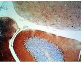
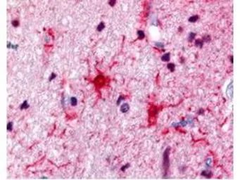
Anti-S100 antibody used in IHC (Paraffin sections) (IHC-P). GTX48819
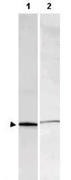
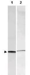
Anti-S100 antibody used in Western Blot (WB). GTX48819

Cat. No. GTX48819

Host

Rabbit

Clonality

Polyclonal

Isotype

IgG

Application

WB, IHC-P, ELISA

Reactivity

Human, Mouse, Rat, Bovine
Package
100 μg ($399)

APPLICATION

Application Note

*Optimal dilutions/concentrations should be determined by the researcher.
Application Recommended Dilution
WB 1:500 - 1:3,000
IHC-P 1:200 - 1:2,000
ELISA 1:5,000 - 1:25,000
Not tested in other applications.

PROPERTIES

Form

Liquid

Buffer

0.02M Potassium Phosphate, 0.15M NaCl

Preservative

0.01% Sodium azide

Storage

Store as concentrated solution. Centrifuge briefly prior to opening vial. For short-term storage (1-2 weeks), store at 4ºC. For long-term storage, aliquot and store at -20ºC or below. Avoid multiple freeze-thaw cycles.

Concentration

5 mg/ml (Please refer to the vial label for the specific concentration.)

Antigen Species

Bovine

Immunogen

This Protein A purified antibody was prepared from whole rabbit serum produced by repeated immunizations with full-length bovine S100 protein (mixture of aa homodimers and ab heterodimers).

Purification

Protein A purified

Conjugation

Unconjugated

RRID

AB_11179831

Note

For laboratory research use only. Not for any clinical, therapeutic, or diagnostic use in humans or animals. Not for animal or human consumption.

Purchasers shall not, and agree not to enable third parties to, analyze, copy, reverse engineer or otherwise attempt to determine the structure or sequence of the product.

TARGET

Synonyms

protein S100-A1

Background

S-100 protein derived from brain tissue is an acidic calcium-binding protein with molecular weight of about 21kDa. In human brain tissue S-100 protein is mainly presented as two isoforms - bb homodimer (S-100b) and ab heterodimer (S-100a). Because of its predominant location in astroglial cells S-100 protein can be used as a sensitive and reliable marker for central nervous system injury. Structural damage of glial cells causes leakage of S-100 protein into the extracellular matrix and into cerebrospinal fluid, further releasing into the bloodstream. Measurements of S-100 protein in patient serum samples are useful in monitoring of traumatic brain injury, ischemic brain damage after circulatory arrests, and in diagnosis and prognosis of clinical outcome in acute stroke. Although predominant among the water-soluble brain proteins, S-100 is also found in a variety of other tissues. S-100 is an intracellular protein that weakly binds calcium. It binds zinc very tightly, however, and this appears to increase the affinity of the protein for calcium. Distinct binding sites, with different affinities, exist for both ions on each monomer. Physiological concentrations of potassium ion antagonize the binding of both divalent cations, especially affecting high-affinity calcium-binding sites.

Database

Research Area

DATA IMAGES

Anti-S100 antibody used in IHC (Paraffin sections) (IHC-P). GTX48819

GTX48819 IHC-P Image

Rabbit anti-S100 was used at a 1:500 dilution to detect S100 by immunohistochemistry in human brain astrocyte tumor tissue. Tissue was formalin-fixed and paraffin embedded.

Anti-S100 antibody used in IHC (Paraffin sections) (IHC-P). GTX48819

GTX48819 IHC-P Image

Rabbit anti-S-100 protein was used at a 1:500 dilution to detect S-100 by immunohistochemistry using a 2-step indirect method. Dark nuclear staining is observed within basket cells located near the Purkinje cells in the cerebellum. Mouse brain tissue was immersed for 24 hours in 10% neutral buffered formalin and paraffin processed followed by sectioning at 4 microns. No antigen unmasking (HIER) or protease digestion was performed prior to immunostaining. Sections were deparaffinized in xylene, and hydrated through graded alcohol to distilled water. All incubations were done at room temperature. All rinses were either distilled water or Tris-HCl with 0.05% Tween 20. Endogenous peroxidase activity was blocked with 3% Hydrogen peroxide for 10'. Primary antibody was diluted as stated and reacted for 30' followed by washes and the addition of donkey anti-rabbit HRP diluted 1:500 for 30'. DAB+ (Dakocytomation) was used as a substrate and was allowed to react for 5'.

Anti-S100 antibody used in Western Blot (WB). GTX48819

GTX48819 WB Image

Western blot using GeneTex's Affinity Purified anti-S-100 antibody shows detection of a band ~11 kDa corresponding to bovine S-100 monomer (100 μg loaded, arrowhead lane 1). The antibody also detects S-100 from rat brain lysate (lane 2). Approximately 35 μg of a rat brain whole cell lysate was separated by 16% SDS-PAGE and transferred onto nitrocellulose. After blocking, the membrane was probed with the primary antibody diluted to 1:1,000 for 2h at room temperature followed by washes and reaction with a 1:10,000 dilution of IRDye™800 conjugated goat anti-Rabbit IgG [H&L] for 45 min at room temperature. IRDye™800 fluorescence image was captured using the Odyssey® Infrared Imaging System developed by LI-COR. IRDye is a trademark of LI-COR, Inc. Other detection systems will yield similar results.

REFERENCE

REVIEW

There are currently no reviews for S100 antibody (GTX48819). Be the first to share your experience with this product.
SDS
Sodium Azide.pdf
Package List Price ($)
$ 399