S100 antibody
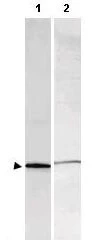
WB analysis of various samples using GTX48819 S100 antibody.
Lane 1 : Bovine S100 protein (100 ng)
Lane 2 : Rat brain (35 μg)
Dilution : 1:1000
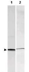
Western blot using GeneTex's Affinity Purified anti-S-100 antibody shows detection of a band ~11 kDa corresponding to bovine S-100 monomer (100 μg loaded, arrowhead lane 1). The antibody also detects S-100 from rat brain lysate (lane 2). Approximately 35 μg of a rat brain whole cell lysate was separated by 16% SDS-PAGE and transferred onto nitrocellulose. After blocking, the membrane was probed with the primary antibody diluted to 1:1,000 for 2h at room temperature followed by washes and reaction with a 1:10,000 dilution of IRDye™800 conjugated goat anti-Rabbit IgG [H&L] for 45 min at room temperature. IRDye™800 fluorescence image was captured using the Odyssey® Infrared Imaging System developed by LI-COR. IRDye is a trademark of LI-COR, Inc. Other detection systems will yield similar results.
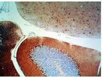
Rabbit anti-S-100 protein was used at a 1:500 dilution to detect S-100 by immunohistochemistry using a 2-step indirect method. Dark nuclear staining is observed within basket cells located near the Purkinje cells in the cerebellum. Mouse brain tissue was immersed for 24 hours in 10% neutral buffered formalin and paraffin processed followed by sectioning at 4 microns. No antigen unmasking (HIER) or protease digestion was performed prior to immunostaining. Sections were deparaffinized in xylene, and hydrated through graded alcohol to distilled water. All incubations were done at room temperature. All rinses were either distilled water or Tris-HCl with 0.05% Tween 20. Endogenous peroxidase activity was blocked with 3% Hydrogen peroxide for 10'. Primary antibody was diluted as stated and reacted for 30' followed by washes and the addition of donkey anti-rabbit HRP diluted 1:500 for 30'. DAB+ (Dakocytomation) was used as a substrate and was allowed to react for 5'.
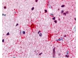
Rabbit anti-S100 was used at a 1:500 dilution to detect S100 by immunohistochemistry in human brain astrocyte tumor tissue. Tissue was formalin-fixed and paraffin embedded.
-
HostRabbit
-
ClonalityPolyclonal
-
IsotypeIgG
-
ApplicationsWB IHC-P ELISA
-
ReactivityHuman, Mouse, Rat, Bovine