SDHA antibody


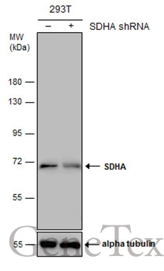
Non-transfected (–) and transfected (+) 293T whole cell extracts (30 μg) were separated by 7.5% SDS-PAGE, and the membrane was blotted with SDHA antibody (GTX101689) diluted at 1:500. The HRP-conjugated anti-rabbit IgG antibody (GTX213110-01) was used to detect the primary antibody.
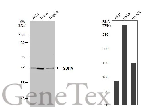
Various whole cell extracts (30 μg) were separated by 7.5% SDS-PAGE, and the membrane was blotted with SDHA antibody (GTX101689) diluted at 1:500. The HRP-conjugated anti-rabbit IgG antibody (GTX213110-01) was used to detect the primary antibody. Corresponding RNA expression data for the same cell lines are based on Human Protein Atlas program.
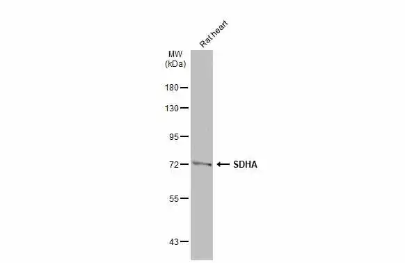
Rat tissue extract (50 μg) was separated by 7.5% SDS-PAGE, and the membrane was blotted with SDHA antibody (GTX101689) diluted at 1:1000. The HRP-conjugated anti-rabbit IgG antibody (GTX213110-01) was used to detect the primary antibody.
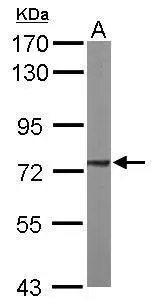
Sample (50 μg of whole cell lysate)
A: mouse brain
7.5% SDS PAGE
GTX101689 diluted at 1:1000
The HRP-conjugated anti-rabbit IgG antibody (GTX213110-01) was used to detect the primary antibody.
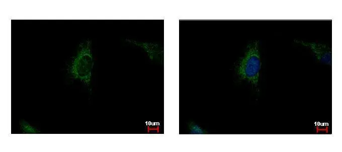
SDHA antibody detects SDHA protein at mitochondria by immunofluorescent analysis.
Sample: HeLa cells were fixed in 4% paraformaldehyde at RT for 15 min.
Green: SDHA protein stained by SDHA antibody (GTX101689) diluted at 1:500.
Blue: Hoechst 33343 staining.
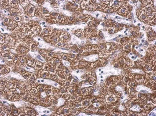
SDHA antibody detects SDHA protein at cytosol on human hepatoma by immunohistochemical analysis.
Sample: Paraffin-embedded hepatoma.
SDHA antibody (GTX101689) dilution: 1:500.
Antigen Retrieval: Trilogy™ (EDTA based, pH 8.0) buffer, 15min
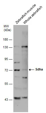
Various tissue extracts (30 μg) were separated by 7.5% SDS-PAGE, and the membrane was blotted with SDHA antibody (GTX101689) diluted at 1:500.
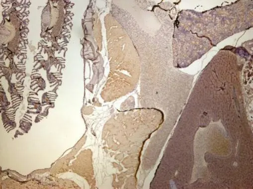
Immunohistochemical analysis of paraffin-embedded zebrafish tissue, using SDHA antibody (GTX101689) at 1:300 dilution.
-
HostRabbit
-
ClonalityPolyclonal
-
IsotypeIgG
-
ApplicationsWB ICC/IF IHC-P
-
ReactivityHuman, Mouse, Rat, Zebrafish