SHP1 antibody

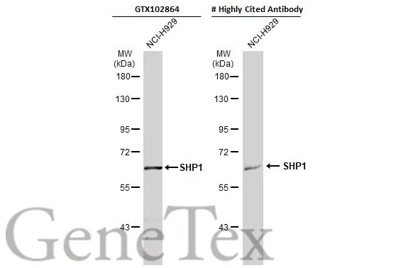
Whole cell extract (30 μg) was separated by 7.5% SDS-PAGE, and the membranes were blotted with SHP1 antibody (GTX102864) diluted at 1:1000 and competitor's antibody (# Highly Cited Antibody) diluted at 1:500. The HRP-conjugated anti-rabbit IgG antibody (GTX213110-01) was used to detect the primary antibody.
*The competitor is not affiliated with GeneTex and does not endorse this product.
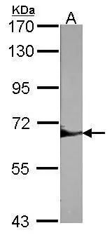
Sample (30 μg of whole cell lysate)
A:NIH-3T3
7.5% SDS PAGE
GTX102864 diluted at 1:1000
The HRP-conjugated anti-rabbit IgG antibody (GTX213110-01) was used to detect the primary antibody.
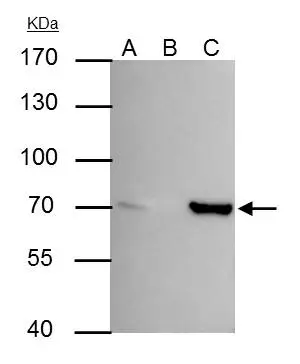
SHP1 antibody immunoprecipitates SHP1 protein in IP experiments. IP Sample: HeLa whole cell lysate/extract A : 30 μg whole cell lysate/extract of SHP1 protein expressing HeLa cells B : Control with 3 μg of pre-immune rabbit IgG C : Immunoprecipitation of SHP1 by 3 μg of SHP1 antibody (GTX102864) 7.5% SDS-PAGE The immunoprecipitated SHP1 protein was detected by SHP1 antibody (GTX102864) diluted at 1 : 500. EasyBlot anti-rabbit IgG (HRP) (GTX221666-01) was used as a secondary reagent.
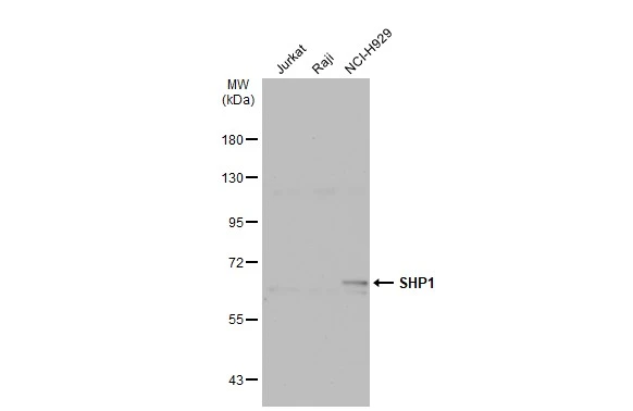
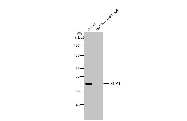
Various whole cell extracts (30 μg) were separated by 7.5% SDS-PAGE, and the membrane was blotted with SHP1 antibody (GTX102864) diluted at 1:1000. The HRP-conjugated anti-rabbit IgG antibody (GTX213110-01) was used to detect the primary antibody.
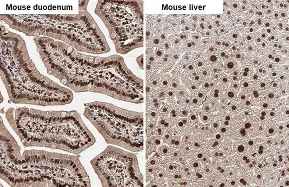
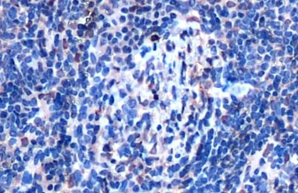
SHP1 antibody detects SHP1 protein by immunohistochemical analysis.Sample: Paraffin-embedded mouse tissues.SHP1 stained by SHP1 antibody (GTX102864) diluted at 1:500.Antigen Retrieval: Citrate buffer, pH 6.0, 15 min
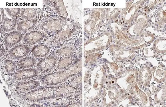
SHP1 antibody detects SHP1 protein by immunohistochemical analysis.Sample: Paraffin-embedded rat tissues.SHP1 stained by SHP1 antibody (GTX102864) diluted at 1:500.Antigen Retrieval: Citrate buffer, pH 6.0, 15 min
Various whole cell extracts (10 μg) were separated by 7.5% SDS-PAGE, and the membrane was blotted with SHP1 antibody (GTX102865) diluted at 1:1000. The HRP-conjugated anti-rabbit IgG antibody (GTX213110-01) was used to detect the primary antibody.
SHP1 antibody detects SHP1 protein at cytoplasm and nucleus by immunohistochemical analysis.Sample: Paraffin-embedded rat liver.SHP1 stained by SHP1 antibody (GTX102864) diluted at 1:500.Antigen Retrieval: Citrate buffer, pH 6.0, 15 min
SHP1 antibody detects SHP1 protein at cytoplasm and nucleus by immunohistochemical analysis.Sample: Paraffin-embedded mouse liver.SHP1 stained by SHP1 antibody (GTX102864) diluted at 1:500.Antigen Retrieval: Citrate buffer, pH 6.0, 15 min
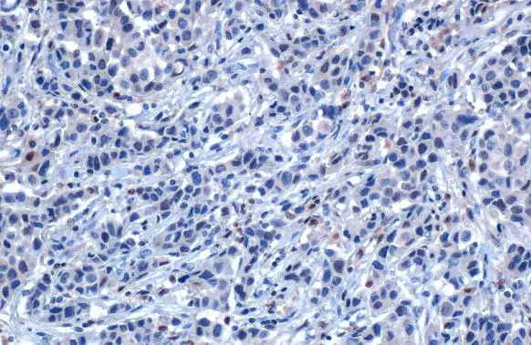
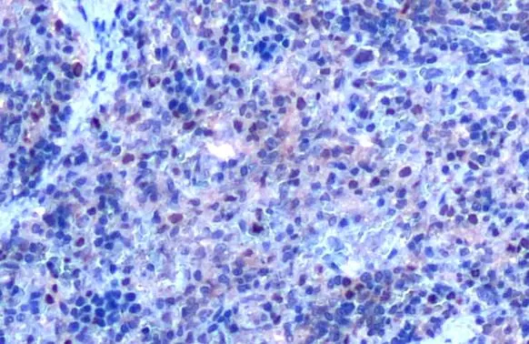
SHP1 antibody detects SHP1 protein at cytoplasm and nucleus by immunohistochemical analysis.Sample: Paraffin-embedded human breast carcinoma.SHP1 stained by SHP1 antibody (GTX102864) diluted at 1:500.Antigen Retrieval: Citrate buffer, pH 6.0, 15 min
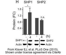
The data was published in the journal PLoS One in 2015. PMID: 25897665
-
HostRabbit
-
ClonalityPolyclonal
-
IsotypeIgG
-
ApplicationsWB IHC-P IHC-Fr IP
-
ReactivityHuman, Mouse, Rat