SP2 antibody


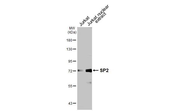
Jurkat whole cell and nuclear extracts (30 μg) were separated by 7.5% SDS-PAGE, and the membrane was blotted with SP2 antibody (GTX129030) diluted at 1:1000. The HRP-conjugated anti-rabbit IgG antibody (GTX213110-01) was used to detect the primary antibody.
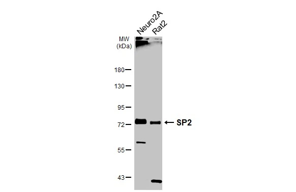
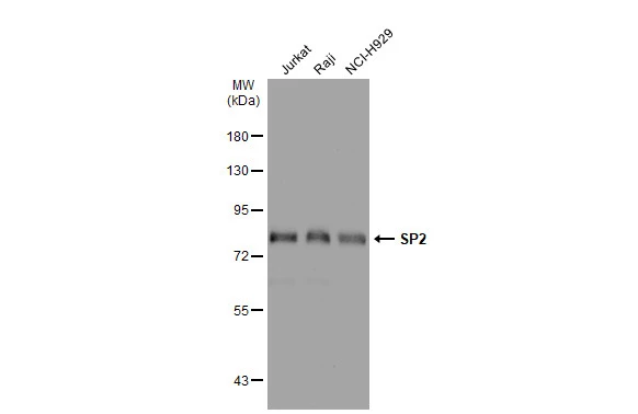
Various whole cell extracts (30 μg) were separated by 7.5% SDS-PAGE, and the membrane was blotted with SP2 antibody (GTX129030) diluted at 1:1000. The HRP-conjugated anti-rabbit IgG antibody (GTX213110-01) was used to detect the primary antibody.
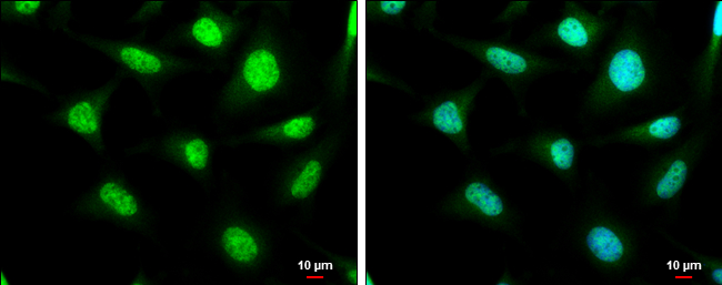
SP2 antibody detects SP2 protein at cytoplasm and nucleus by immunofluorescent analysis.Sample: HeLa cells were fixed in 4% paraformaldehyde at RT for 15 min.Green: SP2 stained by SP2 antibody (GTX129030) diluted at 1:1000.Blue: Hoechst 33342 staining.Scale bar= 10μm.
*The competitor is not affiliated with GeneTex and does not endorse this product.
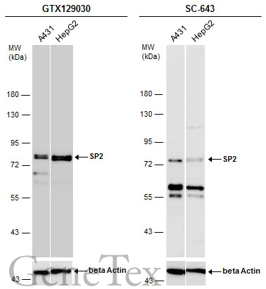
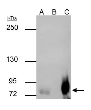
Various whole cell extracts (30 μg) were separated by 7.5% SDS-PAGE, and the membranes were blotted with SP2 antibody (GTX129030) diluted at 1:1000 and competitor's antibody (SC-643) diluted by 1:200.
SP2 antibody immunoprecipitates SP2 protein in IP experiments. IP Sample: Raji whole cell lysate/extract A : 30 μg whole cell lysate/extract of SP2 protein expressing Raji cells B : Control with 3 μg of pre-immune rabbit IgG C : Immunoprecipitation of SP2 by 3 μg of SP2 antibody (GTX129030) 5% SDS-PAGE The immunoprecipitated SP2 protein was detected by SP2 antibody (GTX129030) diluted at 1 : 500. EasyBlot anti-rabbit IgG (HRP) (GTX221666-01) was used as a secondary reagent.
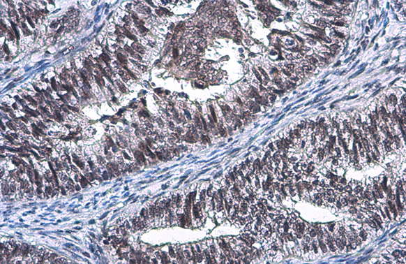
SP2 antibody detects SP2 protein at nucleus by immunohistochemical analysis.Sample: Paraffin-embedded human endometrial carcinoma.SP2 stained by SP2 antibody (GTX129030) diluted at 1:500.Antigen Retrieval: Citrate buffer, pH 6.0, 15 min
Various whole cell extracts (30 μg) were separated by 7.5% SDS-PAGE, and the membrane was blotted with SP2 antibody (GTX129030) diluted at 1:1000. The HRP-conjugated anti-rabbit IgG antibody (GTX213110-01) was used to detect the primary antibody.
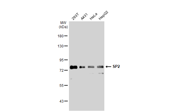
Various whole cell extracts (30 μg) were separated by 7.5% SDS-PAGE, and the membrane was blotted with SP2 antibody (GTX129030) diluted at 1:1000. The HRP-conjugated anti-rabbit IgG antibody (GTX213110-01) was used to detect the primary antibody.
-
HostRabbit
-
ClonalityPolyclonal
-
IsotypeIgG
-
ApplicationsWB ICC/IF IHC-P IP
-
ReactivityHuman, Mouse, Rat