APPLICATION
Application Note
*Optimal dilutions/concentrations should be determined by the researcher.
Application |
Dilution |
1:500-1:3000 |
Not tested in other applications.
Calculated MW
Positive Control
A549 , A549 membrane extract , HepG2 , HepG2 cytoplasmic extract
Predict Reactivity
Mouse, Rat, Bovine, Pig(>80% identity)
PROPERTIES
Form
Liquid
Buffer
1XPBS, 20% Glycerol (pH7). 0.025% ProClin 300 was added as a preservative.
Storage
Store as concentrated solution. Centrifuge briefly prior to opening vial. For short-term storage (1-2 weeks), store at 4ºC. For long-term storage, aliquot and store at -20ºC or below. Avoid multiple freeze-thaw cycles.
Concentration
2.39mg/ml(Please refer to the vial label for the specific concentration.)
Antigen Species
Human
Immunogen
Carrier-protein conjugated synthetic peptide encompassing a sequence within the center region of human TGF beta Receptor I. The exact sequence is proprietary.
Purification
Purified by antigen-affinity chromatography.
Conjugation
Unconjugated
Note
For laboratory use only. Not for any clinical, therapeutic, or diagnostic use in humans or animals. Not for animal or human consumption.
TARGET
Synonyms
AAT5 antibody , ACVRLK4 antibody , ALK5 antibody , LDS1A antibody , LDS2A antibody , MSSE antibody , SKR4 antibody , TGFBR1 antibody , TGFR-1 antibody , TGFR1 antibody , transforming growth factor beta receptor I antibody
Cellular Localization
Membrane; Single-pass type I membrane protein
Background
The protein encoded by this gene forms a heteromeric complex with type II TGF-beta receptors when bound to TGF-beta, transducing the TGF-beta signal from the cell surface to the cytoplasm. The encoded protein is a serine/threonine protein kinase. Mutations in this gene have been associated with Loeys-Dietz aortic aneurysm syndrome (LDAS). Multiple transcript variants encoding different isoforms have been found for this gene. [provided by RefSeq, Aug 2008]
Database
Research Area
DATA IMAGES
|
GTX134209 WB Image
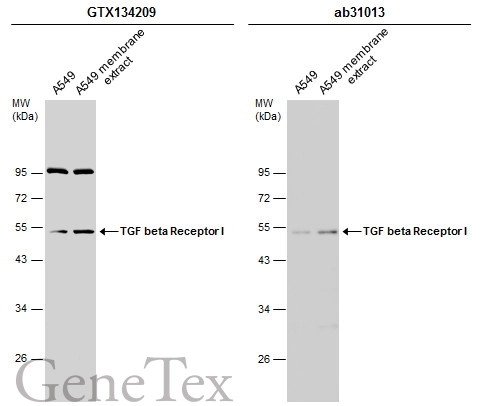
A549 whole cell and membrane extracts (30 µg) were separated by 10% SDS-PAGE, and the membranes were blotted with TGF beta Receptor I antibody (GTX134209) diluted at 1:1000 and competitor's antibody (ab31013) diluted at 1:500. The HRP-conjugated anti-rabbit IgG antibody (GTX213110-01) was used to detect the primary antibody, and the signal was developed with Trident ECL plus-Enhanced.
*The competitor is not affiliated with GeneTex and does not endorse this product.
|
|
GTX134209 WB Image
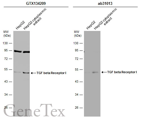
HepG2 whole cell and cytoplasmic extracts (30 µg) were separated by 10% SDS-PAGE, and the membranes were blotted with TGF beta Receptor I antibody (GTX134209) diluted at 1:1000 and competitor's antibody (ab31013) diluted at 1:500. The HRP-conjugated anti-rabbit IgG antibody (GTX213110-01) was used to detect the primary antibody.
*The competitor is not affiliated with GeneTex and does not endorse this product.
|