beta Amyloid antibody
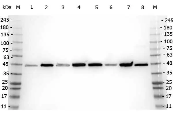
WB analysis of various samples using GTX82974 beta Amyloid antibody.
Lane 1 : 293T whole cell lysate
Lane 2 : HeLa whole cell lysate
Lane 3 : MCF-7 whole cell lysate
Lane 4 : Jurkat whole cell lysate
Lane 5 : A431 whole cell lysate
Lane 6 : LNCaP whole cell lysate
Lane 7 : A-172 whole cell lysate
Lane 8 : NIH-3T3 whole cell lysate
Loading : 35 μg
Dilution : 1:5000
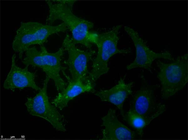
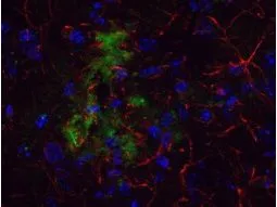
ICC/IF analysis of MeOH-fixed HeLa cells using GTX82974 beta Amyloid antibody.
Green : Primary antibody
Blue : DAPI
APP is a cell surface protein that rapidly becomes internalized to endosomes and lysosomes. Some APP accumulates in secretory transport vesicles. Colocalizes with other proteins in a vesicular pattern in cytoplasm and perinuclear regions.
Dilution : 1 μg/mL
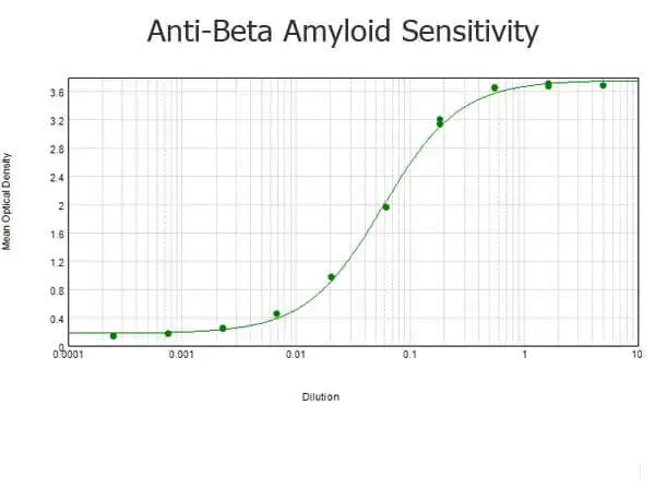
ELISA analysis of BSA-conjugated immunizing peptide using serially diluted GTX82974 beta Amyloid antibody.
Coating : 0.1 μg
WB analysis of various samples using GTX82974 beta Amyloid antibody.
Lane 1 : Protein ladder
Lane 2 : 293T whole cell lysate
Lane 3 : Mouse brain tissus lysate
Lane 4 : A-172 whole cell lysate
Loading : 10 μg
Dilution : 1 μg/mL
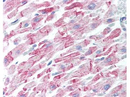
IHC-P analysis of human heart tissue using GTX82974 beta Amyloid antibody.
Dilution : 5 μg/mL
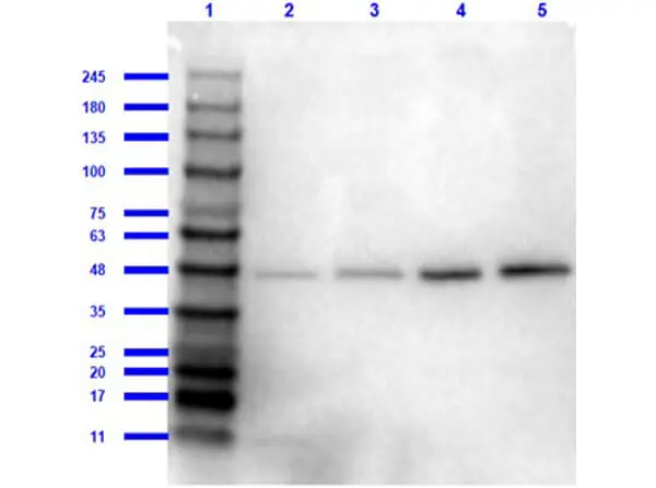
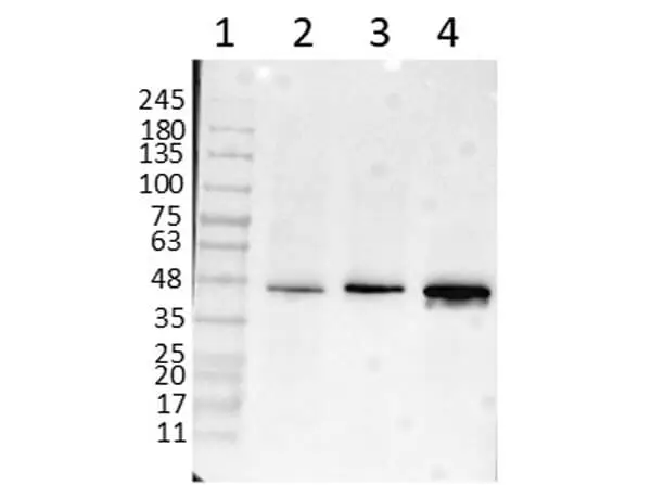
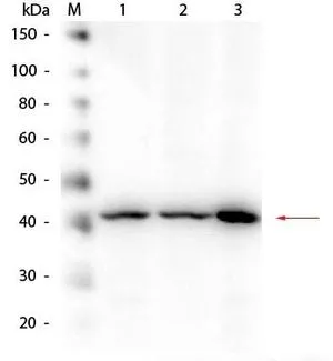
WB analysis of various samples using GTX82974 beta Amyloid antibody.
Lane 1 : Protein ladder
Lane 2 : 293T whole cell lysate
Lane 3 : Mouse brain tissue lysate
Lane 4 : A-172 whole cell lysate
Lane 5 : Daudi whole cell lysate
Loading : 10 μg/mL
Dilution : 1:1000
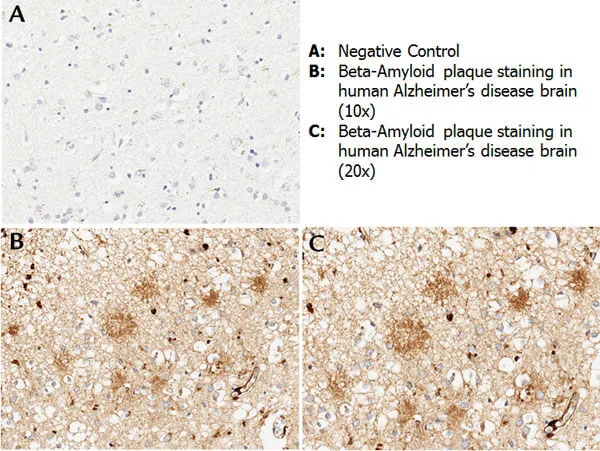
IHC-P analysis of human Alzheimer's disease brain tissue using GTX82974 beta Amyloid antibody.
Antigen retrieval : Heated mediated citrate buffer (pH 6.0)
Dilution : 1:100
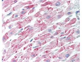
Human Heart (formalin-fixed, paraffin-embedded) stained with Anti-Beta Amyloid Antibody (GTX82974) at 5 ug/ml followed by biotinylated goat anti-rabbit IgG secondary antibody, alkaline phosphatase-streptavidin and chromogen.
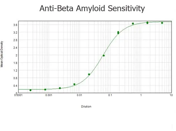
ELISA results of GTX82974 beta Amyloid antibody tested against BSA-conjugated peptide of immunizing peptide. Each well was coated in duplicate with 0.1μg of conjugate. The starting dilution of antibody was 5μg/ml and the X-axis represents the Log10 of a 3-fold dilution. This titration is a 4-parameter curve fit where the IC50 is defined as the titer of the antibody. Assay performed using 3% fish gel, Goat anti-Rabbit IgG Antibody Peroxidase Conjugated and TMB Substrate.
Immunohistochemistical detection of beta Amyloid using Anti-Beta Amyloid Antibody on TG APP23 mouse brain cortex frozen sections. Anti-Beta Amyloid Antibody used at 1/200 and incubated for 2 hours in TBS/BSA/Tween/azide. Fluorescent labelled anti rabbit IgG was then added.
Western Blot of Rabbit anti-Beta Amyloid Antibody. Lane 1: HEK293 WCL. Lane 2: Mouse Brain WCL. Lane 3: A-172 WCL. Load: 10.0 μg per lane. Primary antibody: Beta Amyloid Antibody at 1:1,000 overnight at 4ºC. Secondary antibody: Peroxidase Conjugated Goat-a-Rabbit IgG at 1:40,000 for 30 min at RT. Block for 30 min at RT. Predicted/Observed size: 40 kDa, 40 kDa for Beta Amyloid.
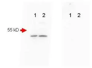
Mouse Brain (Lane 1) and Mouse Spinal Chord (Lane 2) were run on a 4-20% gradient gel, Blocked in 1% BSA-TBS-T 30 min RT and probed with Rb-a-Beta Amyloid (GTX82974) 1:1000 in 1% BSA-TBS-T o/n 4ºC. FEMTOMAX chemiluminescent substrate was used for detection of a 40-50 kD band consistent with a higher MW precursor form of beta amyloid. A secondary Ab only control (Shown right) showed no detectable signal.
-
HostRabbit
-
ClonalityPolyclonal
-
IsotypeIgG
-
ApplicationsWB ICC/IF IHC-P IHC-Fr ELISA
-
ReactivityHuman, Mouse