hnRNP A2B1 antibody

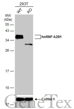
Wild-type (WT) and hnRNP A2B1 knockout (KO) HEK293T cell extracts (30 μg) were separated by 12% SDS-PAGE, and the membrane was blotted with hnRNP A2B1 antibody (GTX127928) diluted at 1:40000. The HRP-conjugated anti-rabbit IgG antibody (GTX213110-01) was used to detect the primary antibody.
hnRNP A2B1 antibody detects hnRNP A2B1 protein at nucleus by immunohistochemical analysis.Sample: Paraffin-embedded mouse intestine.hnRNP A2B1 stained by hnRNP A2B1 antibody (GTX127928) diluted at 1:500.Antigen Retrieval: Citrate buffer, pH 6.0, 15 min
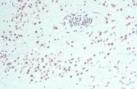
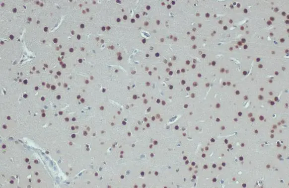
hnRNP A2B1 antibody detects hnRNP A2B1 protein at nucleus by immunohistochemical analysis.
Sample: Paraffin-embedded rat brain.
hnRNP A2B1 stained by hnRNP A2B1 antibody (GTX127928) diluted at 1:500.
Antigen Retrieval: Citrate buffer, pH 6.0, 15 min
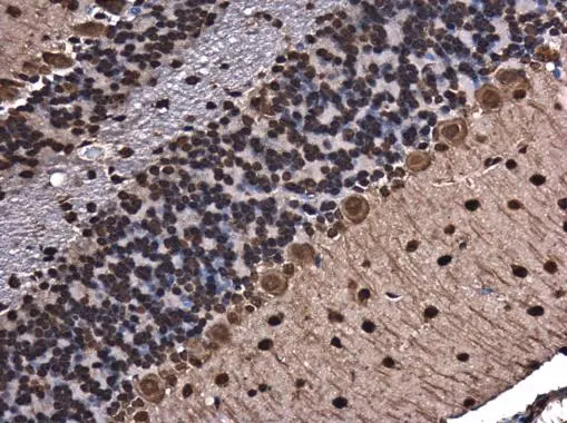
hnRNP A2B1 antibody detects hnRNP A2B1 protein at cytoplasm and nucleus in mouse brain by immunohistochemical analysis.
Sample: Paraffin-embedded mouse brain.
hnRNP A2B1 antibody (GTX127928) diluted at 1:500.
Antigen Retrieval: Citrate buffer, pH 6.0, 15 min
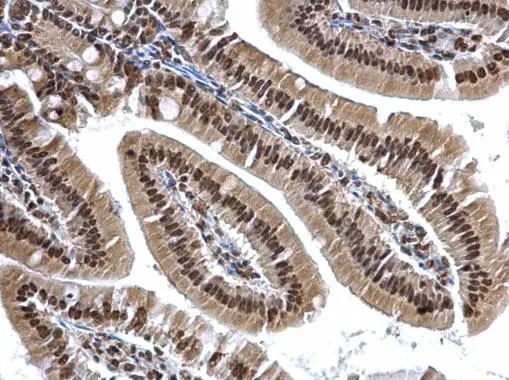
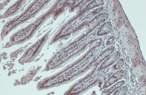
hnRNP A2B1 antibody detects hnRNP A2B1 protein at cytosol and nucleus on mouse duodenum by immunohistochemical analysis.
Sample: Paraffin-embedded mouse duodenum.
hnRNP A2B1 antibody (GTX127928) dilution: 1:500.
Antigen Retrieval: Trilogy™ (EDTA based, pH 8.0) buffer, 15min
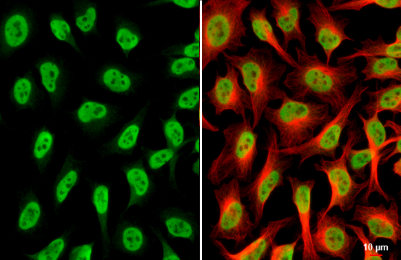
hnRNP A2B1 antibody detects hnRNP A2B1 protein at endoplasmic reticulum and nucleus by immunofluorescent analysis.Sample: HeLa cells were fixed in 4% paraformaldehyde at RT for 15 min.Green: hnRNP A2B1 stained by hnRNP A2B1 antibody (GTX127928) diluted at 1:500.Red: alpha Tubulin, a cytoskeleton marker, stained by alpha Tubulin antibody [GT114] (GTX628802) diluted at 1:1000.Scale bar= 10μm.
hnRNP A2B1 antibody detects hnRNP A2B1 protein at nucleus by immunohistochemical analysis.Sample: Paraffin-embedded mouse brain.hnRNP A2B1 stained by hnRNP A2B1 antibody (GTX127928) diluted at 1:500.Antigen Retrieval: Citrate buffer, pH 6.0, 15 min

hnRNP A2B1 antibody detects hnRNP A2B1 protein by immunohistochemical analysis.Sample: Paraffin-embedded rat tissues.hnRNP A2B1 stained by hnRNP A2B1 antibody (GTX127928) diluted at 1:500.Antigen Retrieval: Citrate buffer, pH 6.0, 15 min
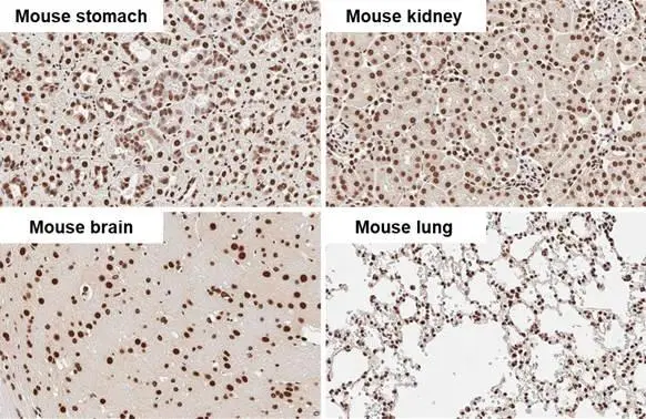
hnRNP A2B1 antibody detects hnRNP A2B1 protein by immunohistochemical analysis.Sample: Paraffin-embedded mouse tissues.hnRNP A2B1 stained by hnRNP A2B1 antibody (GTX127928) diluted at 1:500.Antigen Retrieval: Citrate buffer, pH 6.0, 15 min
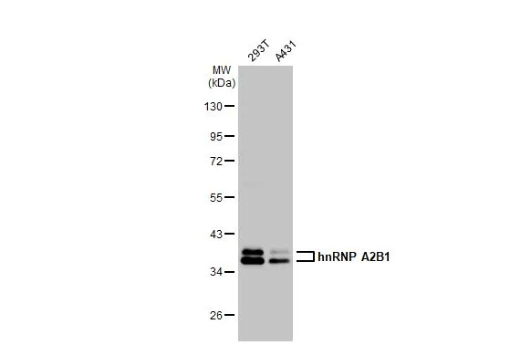
Various whole cell extracts (30 μg) were separated by 10% SDS-PAGE, and the membrane was blotted with hnRNP A2B1 antibody (GTX127928) diluted at 1:10000. The HRP-conjugated anti-rabbit IgG antibody (GTX213110-01) was used to detect the primary antibody.
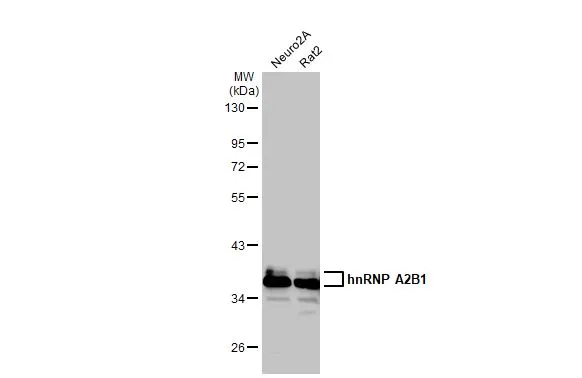
Various whole cell extracts (30 μg) were separated by 10% SDS-PAGE, and the membrane was blotted with hnRNP A2B1 antibody (GTX127928) diluted at 1:10000. The HRP-conjugated anti-rabbit IgG antibody (GTX213110-01) was used to detect the primary antibody.
-
HostRabbit
-
ClonalityPolyclonal
-
IsotypeIgG
-
ApplicationsWB ICC/IF IHC-P
-
ReactivityHuman, Mouse, Rat