Iba1 antibody

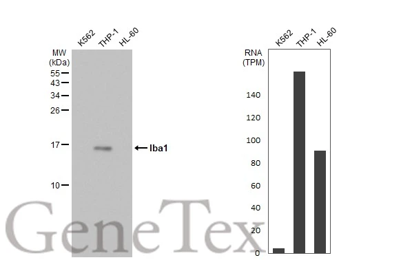
Various whole cell extracts (30 μg) were separated by 15% SDS-PAGE, and the membrane was blotted with Iba1 antibody (GTX100042) diluted at 1:1000. The HRP-conjugated anti-rabbit IgG antibody (GTX213110-01) was used to detect the primary antibody. Corresponding RNA expression data for the same cell lines are based on Human Protein Atlas program.
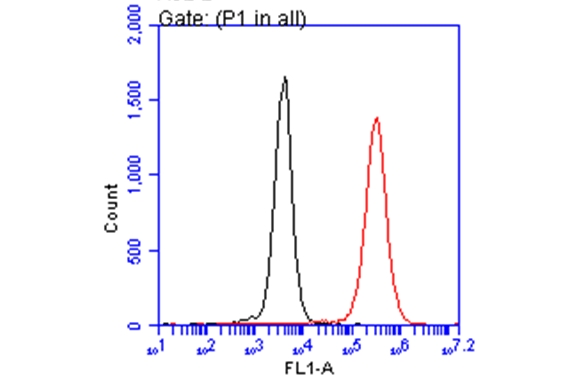
Iba1 antibody (GTX100042) detects AIF1 protein by flow cytometry analysis.
Sample: THP-1 cell.
Black: Unlabelled sample was used as a control.
Red: Iba1 antibody (GTX100042) dilution: 1:50.
Acquisition of 20,000 events were collected for FACS analysis.
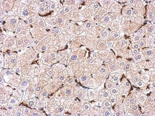
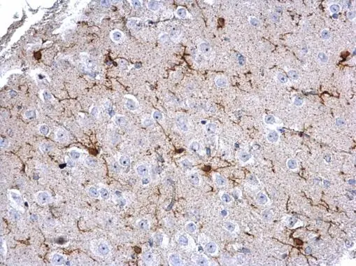
Iba1 antibody detects Iba1 in the cytosol of Kupffer cell in human hepatoma by immunohistochemical analysis.
Immunohistochemical analysis of paraffin-embedded human hepatoma, using Iba1(GTX100042) antibody at 1:500 dilution.
Antigen Retrieval: Trilogy™ (EDTA based, pH 8.0) buffer, 15min
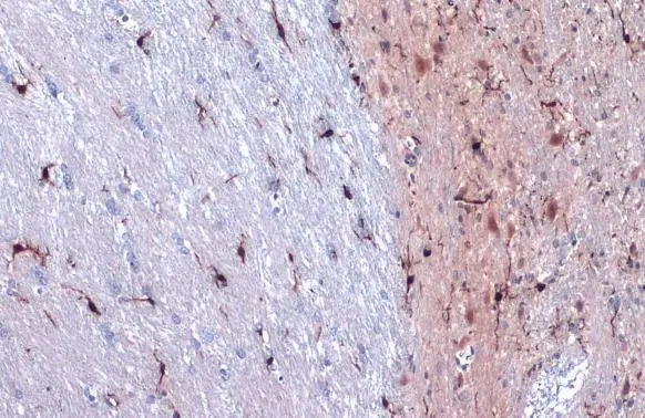
Iba1 antibody detects Iba1 protein at cell membrane and cytoplasm by immunohistochemical analysis.Sample: Paraffin-embedded rat cerebellum.Iba1 stained by Iba1 antibody (GTX100042) diluted at 1:500.Antigen Retrieval: Citrate buffer, pH 6.0, 15 min
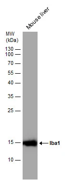
Mouse tissue extract (50 μg) was separated by 15% SDS-PAGE, and the membrane was blotted with Iba1 antibody (GTX100042) diluted at 1:1000.
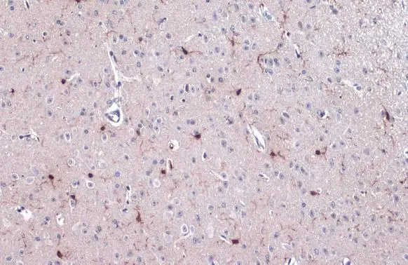
Iba1 antibody detects Iba1 protein on mouse fore brain by immunohistochemical analysis.
Sample: Paraffin-embedded mouse fore brain.
Iba1 antibody (GTX100042) dilution: 1:500.
Antigen Retrieval: Trilogy™ (EDTA based, pH 8.0) buffer, 15min
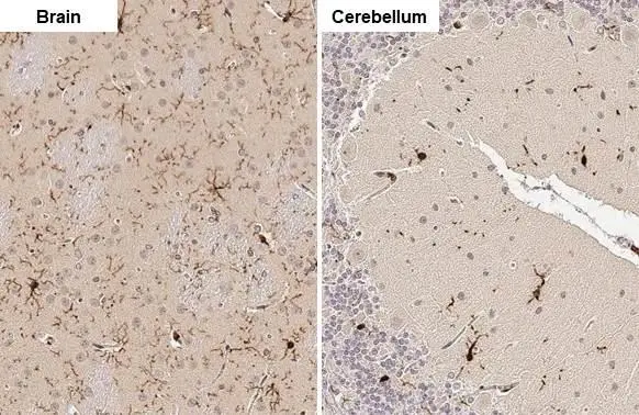
Iba1 antibody detects Iba1 protein at cell membrane and cytoplasm by immunohistochemical analysis.Sample: Paraffin-embedded mouse cerebellum.Iba1 stained by Iba1 antibody (GTX100042) diluted at 1:500.Antigen Retrieval: Citrate buffer, pH 6.0, 15 min
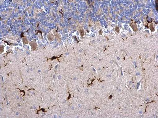
Iba1 antibody detects Iba1 protein on rat hind brain by immunohistochemical analysis.
Sample: Paraffin-embedded rat hind brain.
Iba1 antibody (GTX100042) dilution: 1:500.
Antigen Retrieval: Trilogy™ (EDTA based, pH 8.0) buffer, 15min
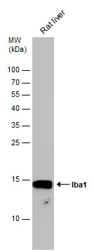
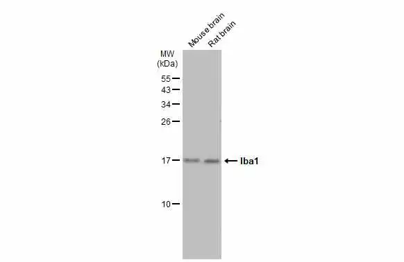
Rat tissue extract (50 μg) was separated by 15% SDS-PAGE, and the membrane was blotted with Iba1 antibody (GTX100042) diluted at 1:1000.
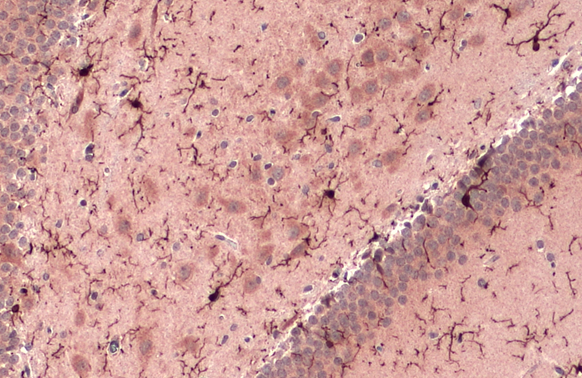
Iba1 antibody detects Iba1 protein at cell membrane and cytoplasm by immunohistochemical analysis.Sample: Paraffin-embedded mouse brain.Iba1 stained by Iba1 antibody (GTX100042) diluted at 1:500.Antigen Retrieval: Citrate buffer, pH 6.0, 15 min
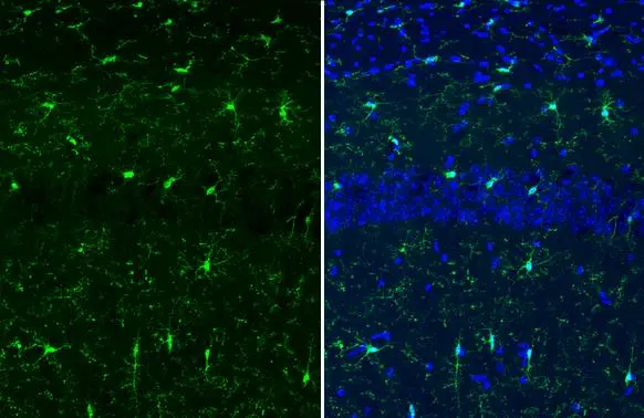
Iba1 antibody detects Iba1 protein at cell membrane and cytoplasm by immunohistochemical analysis.Sample: Frozen-sectioned mouse brain.Green: Iba1 stained by Iba1 antibody (GTX100042) diluted at 1:500.Blue: Fluoroshield with DAPI (GTX30920).Antigen Retrieval: ice-cold MeOH for 5 min
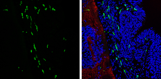
Iba1 antibody detects Iba1 protein expression at microglias by immunohistochemical analysis.
Sample: Frozen sectioned E13.5 Rat brain.
Green: Iba1 protein stained by Iba1 antibody (GTX100042) diluted at 1:250.
Red: beta Tubulin 3/ TUJ1, a mature neuron marker, stained by beta Tubulin 3/ TUJ1 antibody [GT11710] (GTX631836) diluted at 1:500.
Blue: Fluoroshield with DAPI (GTX30920).
Iba1 antibody detects Iba1 protein at cell membrane and cytoplasm by immunohistochemical analysis.Sample: Paraffin-embedded rat brain.Iba1 stained by Iba1 antibody (GTX100042) diluted at 1:500.Antigen Retrieval: Citrate buffer, pH 6.0, 15 min
Various tissue extracts (50 μg) were separated by 15% SDS-PAGE, and the membrane was blotted with Iba1 antibody (GTX100042) diluted at 1:1000. The HRP-conjugated anti-rabbit IgG antibody (GTX213110-01) was used to detect the primary antibody.
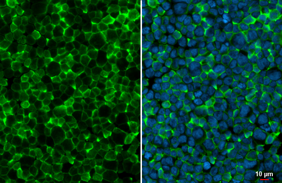
Iba1 antibody detects Iba1 protein at cell membrane by immunofluorescent analysis.Sample: THP-1 cells were fixed in 4% paraformaldehyde at RT for 15 min.Green: Iba1 stained by Iba1 antibody (GTX100042) diluted at 1:500.Blue: Fluoroshield with DAPI (GTX30920).
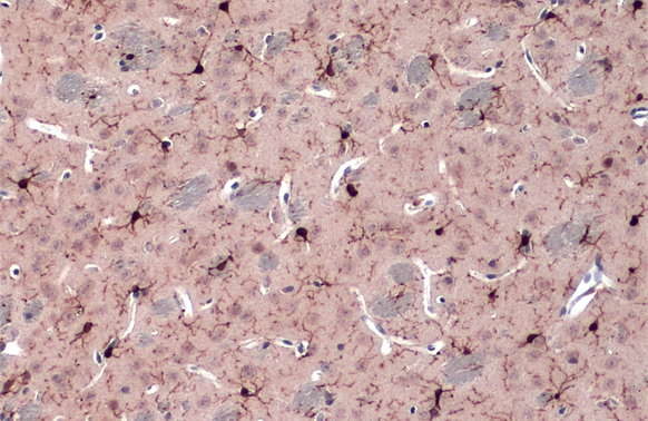
Iba1 antibody detects Iba1 protein by immunohistochemical analysis.Sample: Paraffin-embedded rat tissues.Iba1 stained by Iba1 antibody (GTX100042) diluted at 1:100.Antigen Retrieval: Citrate buffer, pH 6.0, 15 min
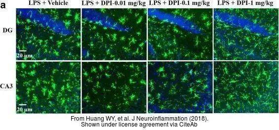
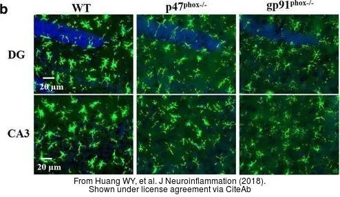
The data was published in the journal J Neuroinflammation in 2018. PMID: 29753328
The data was published in the journal J Neuroinflammation in 2018. PMID: 29753328
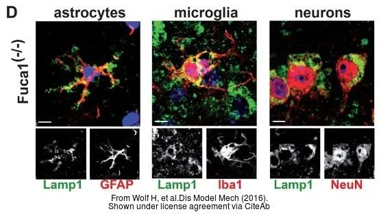
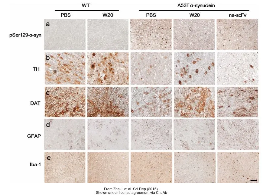
The data was published in the journal Dis Model Mech in 2016. PMID: 27491075
_1580_24041501_559.webp)
The data was published in the journal Front Immunol in 2018.PMID: 29662492
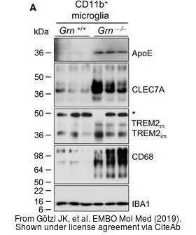
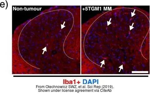
The data was published in the journal EMBO Mol Med in 2019.PMID: 31122931
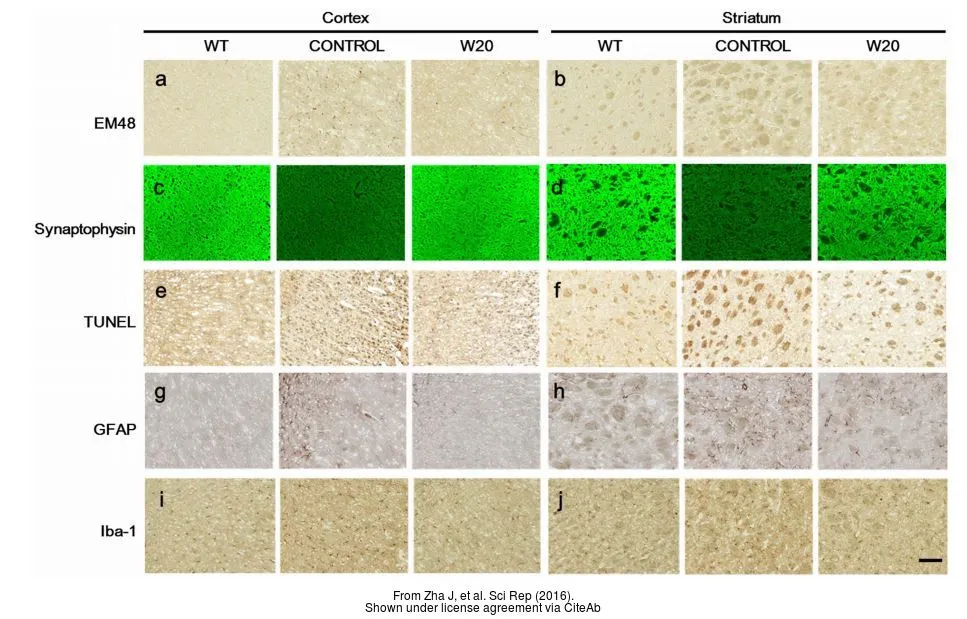
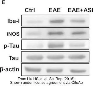
The data was published in the journal Sci Rep in 2016.PMID: 27824125
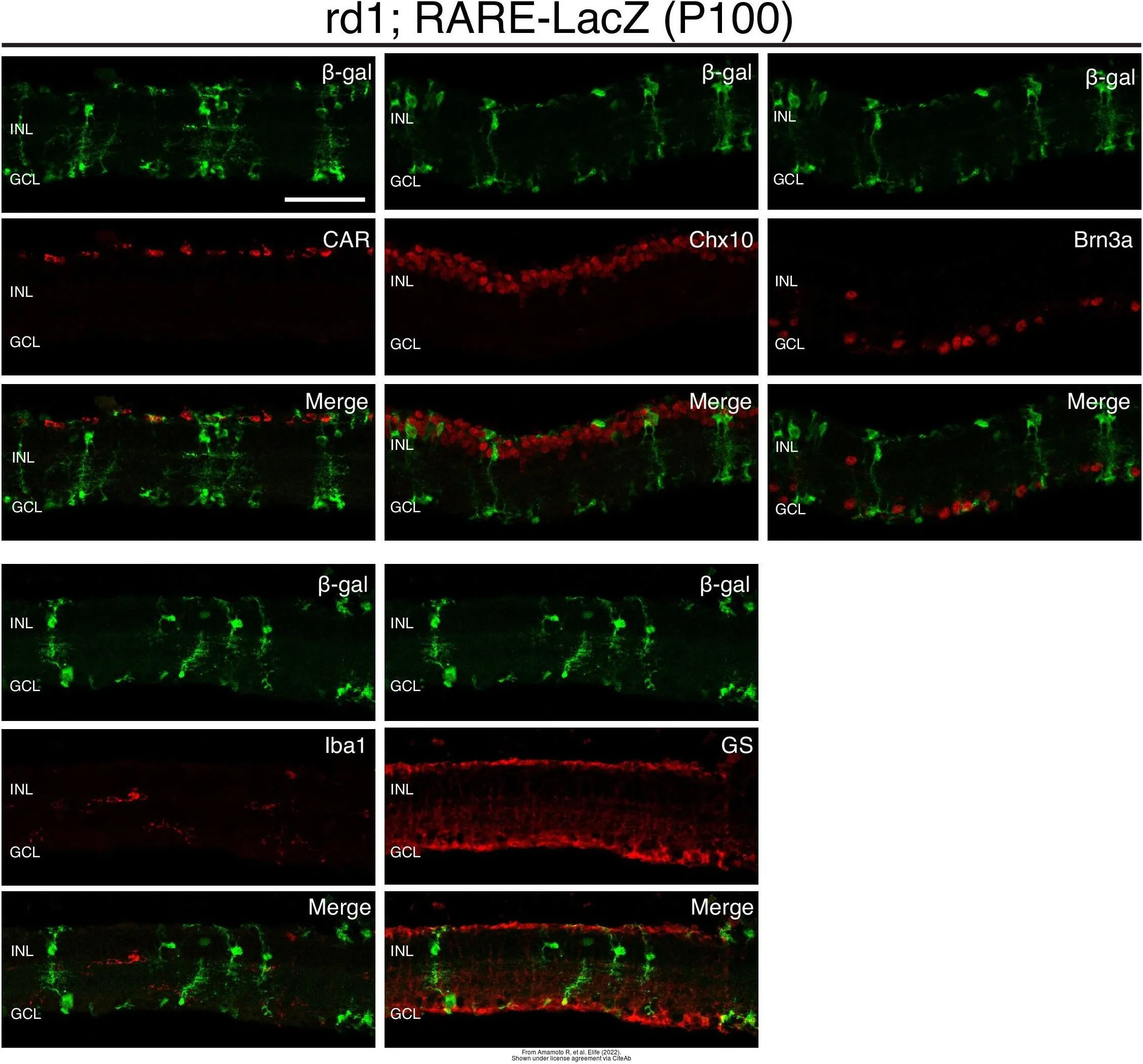
The data was published in the 2022 in Elife. PMID: 35315776
-
HostRabbit
-
ClonalityPolyclonal
-
IsotypeIgG
-
ApplicationsWB ICC/IF IHC-P IHC-Fr FCM IHC IHC (Free Floating)
-
ReactivityHuman, Mouse, Rat