Normal Tissue Gallery- GTX04963

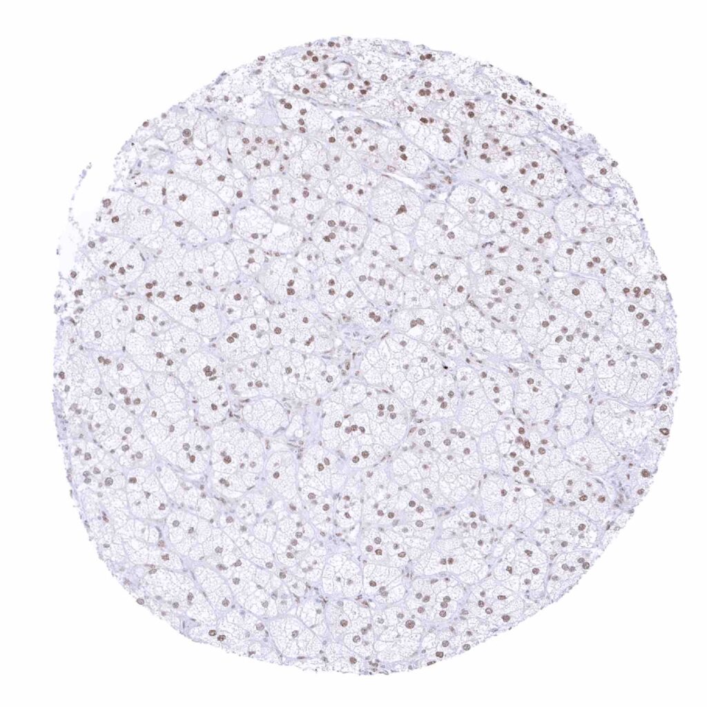
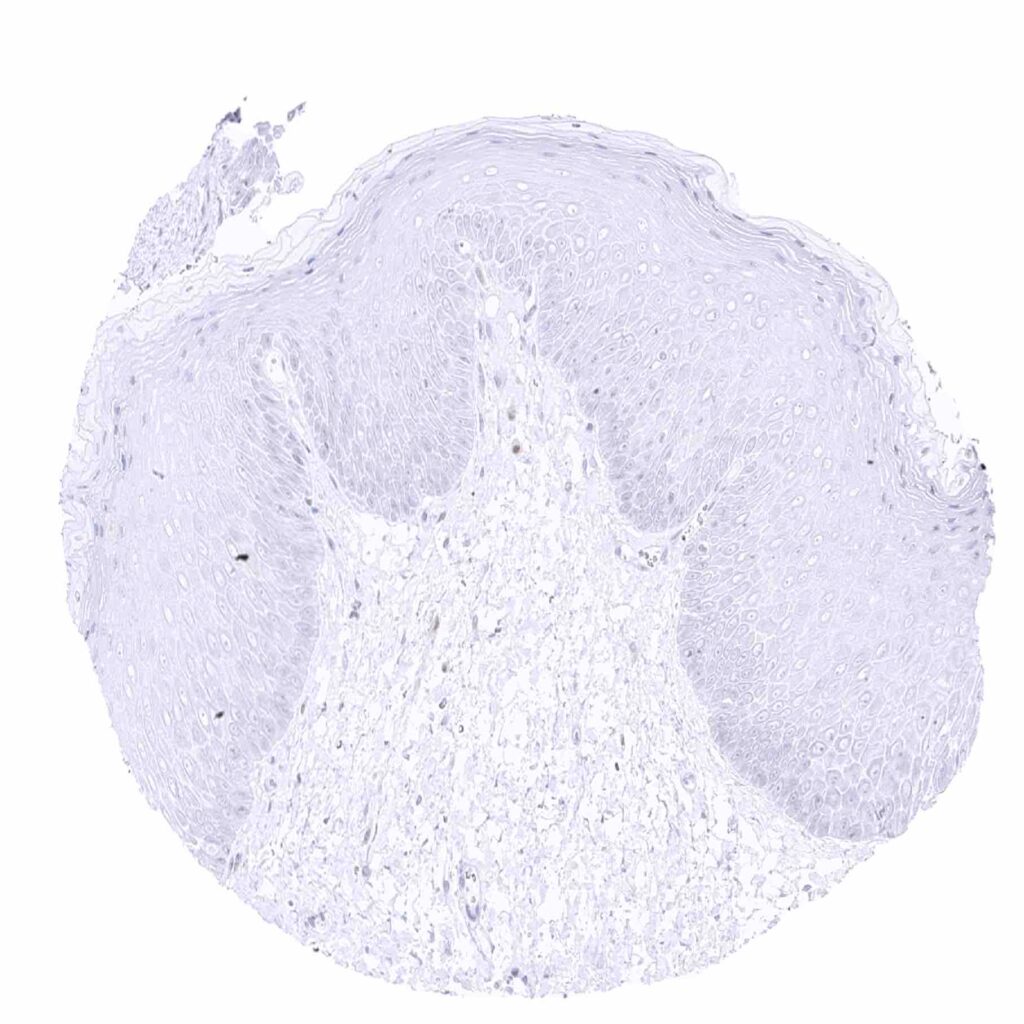
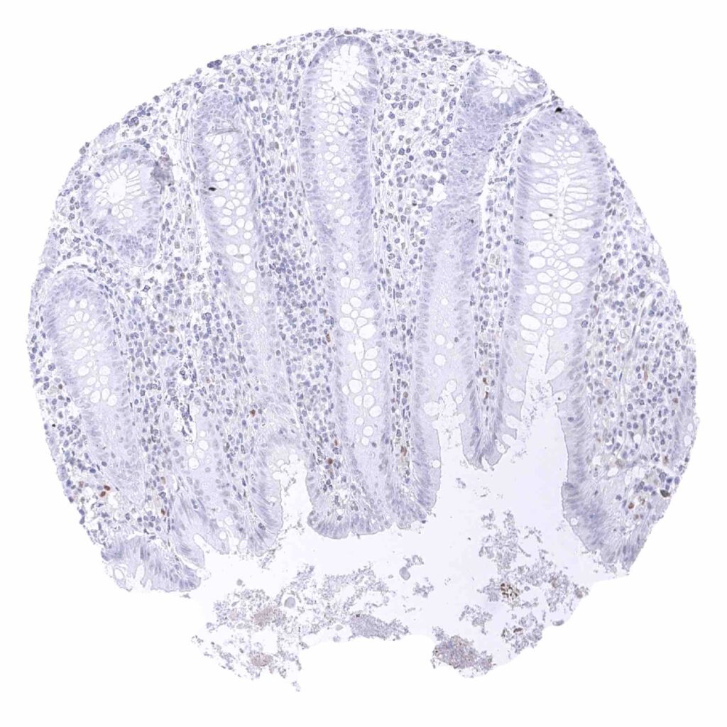
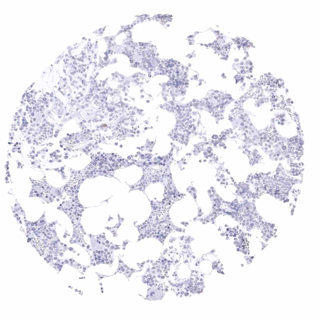
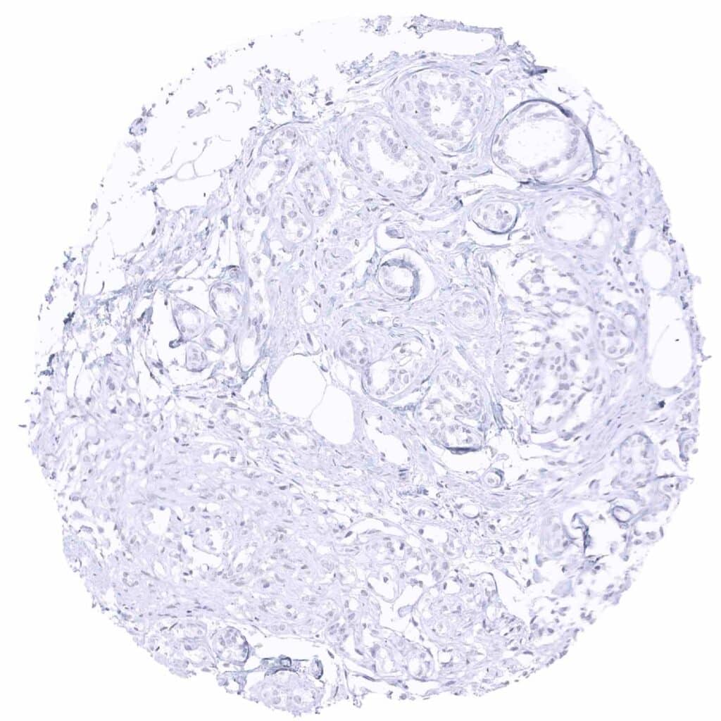
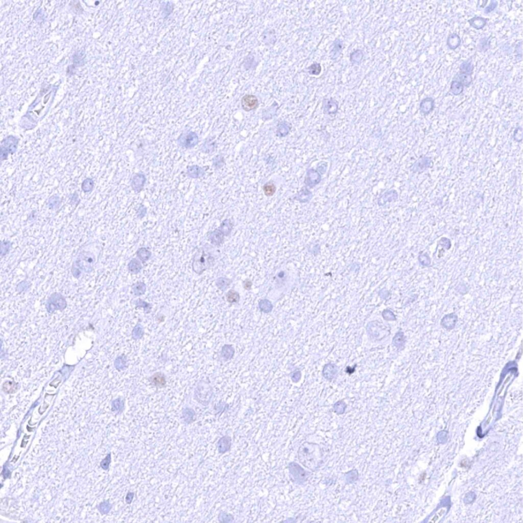
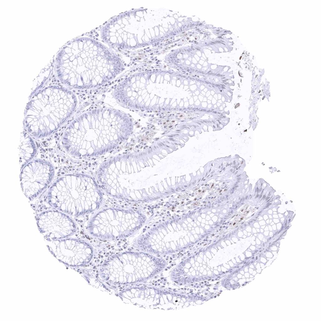
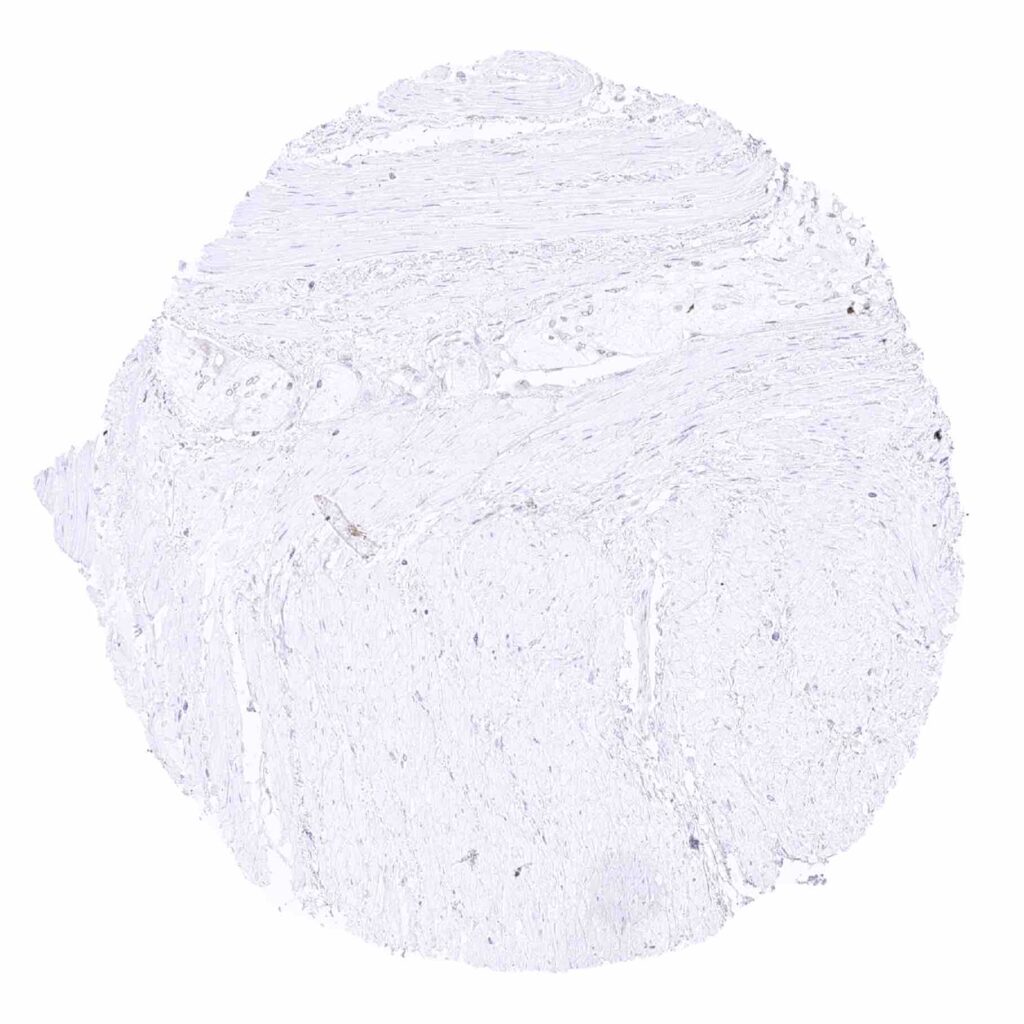
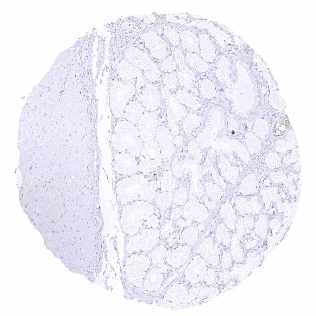
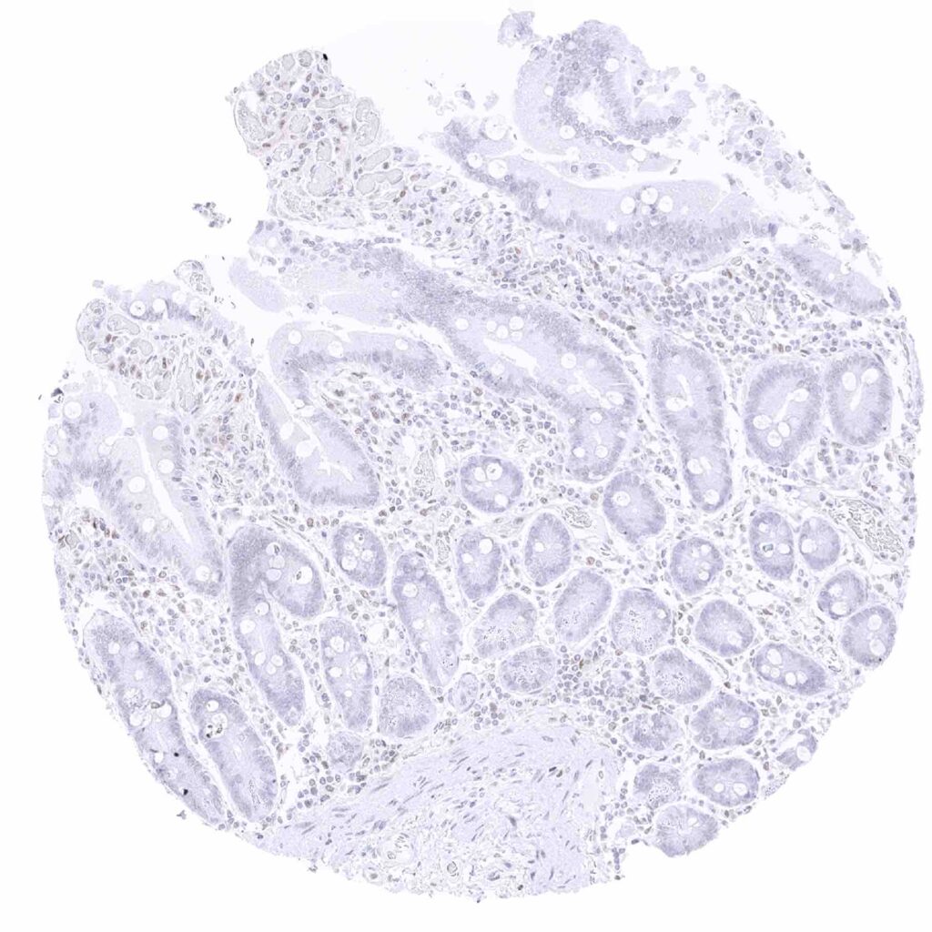
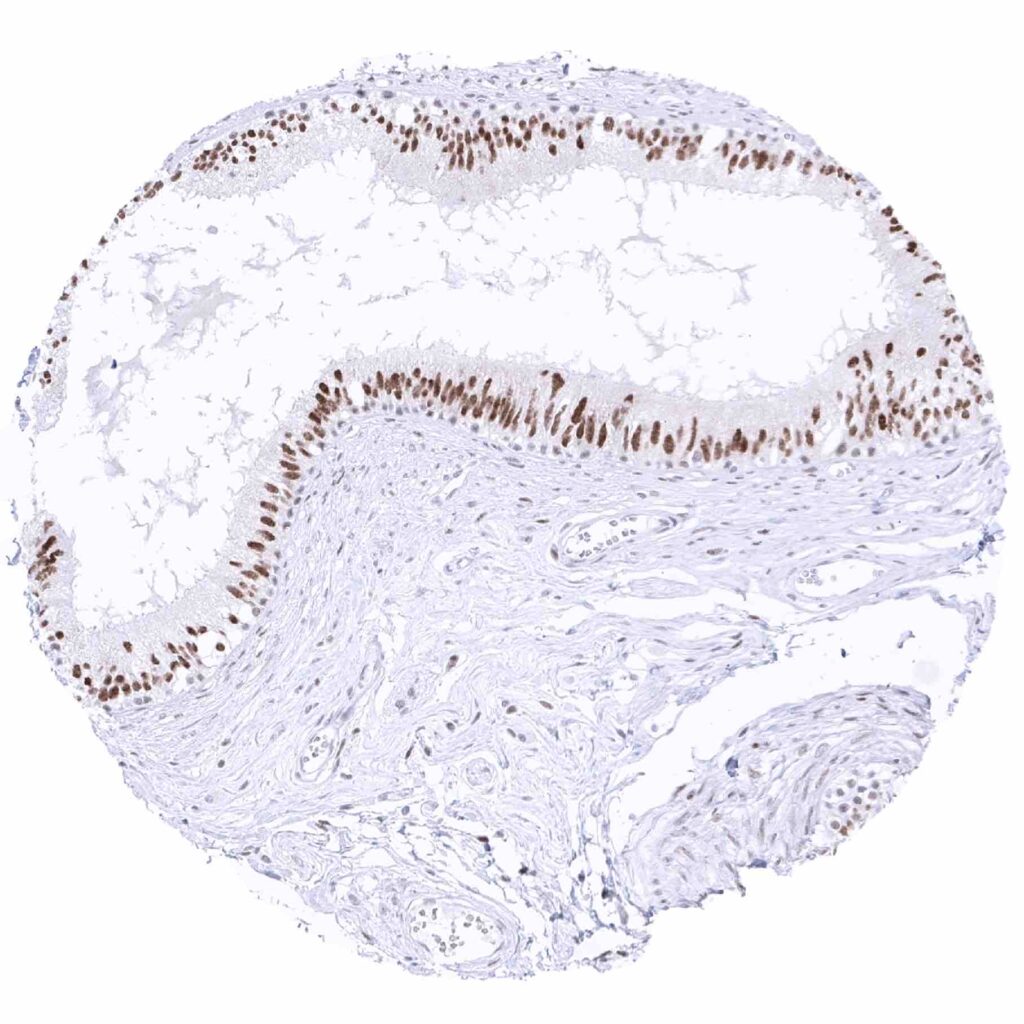
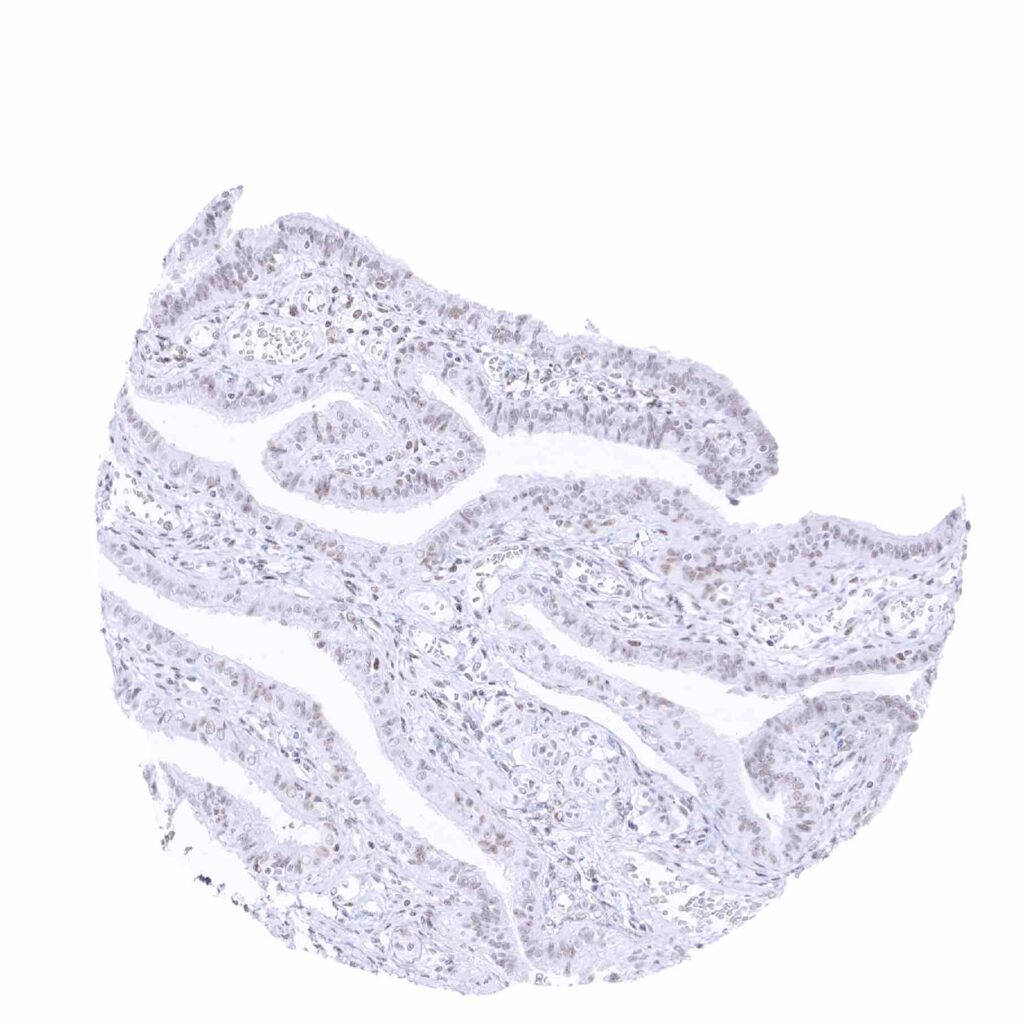
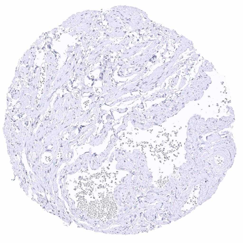
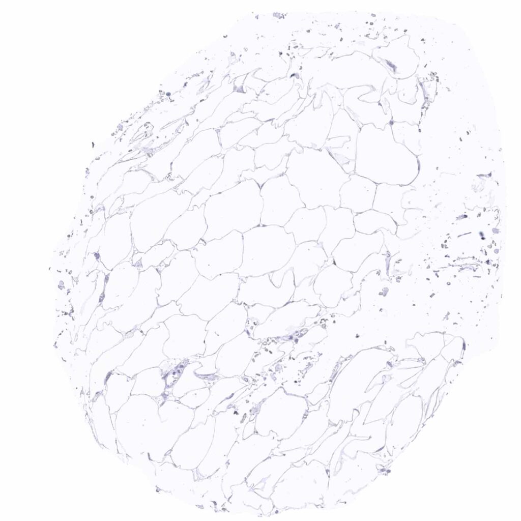
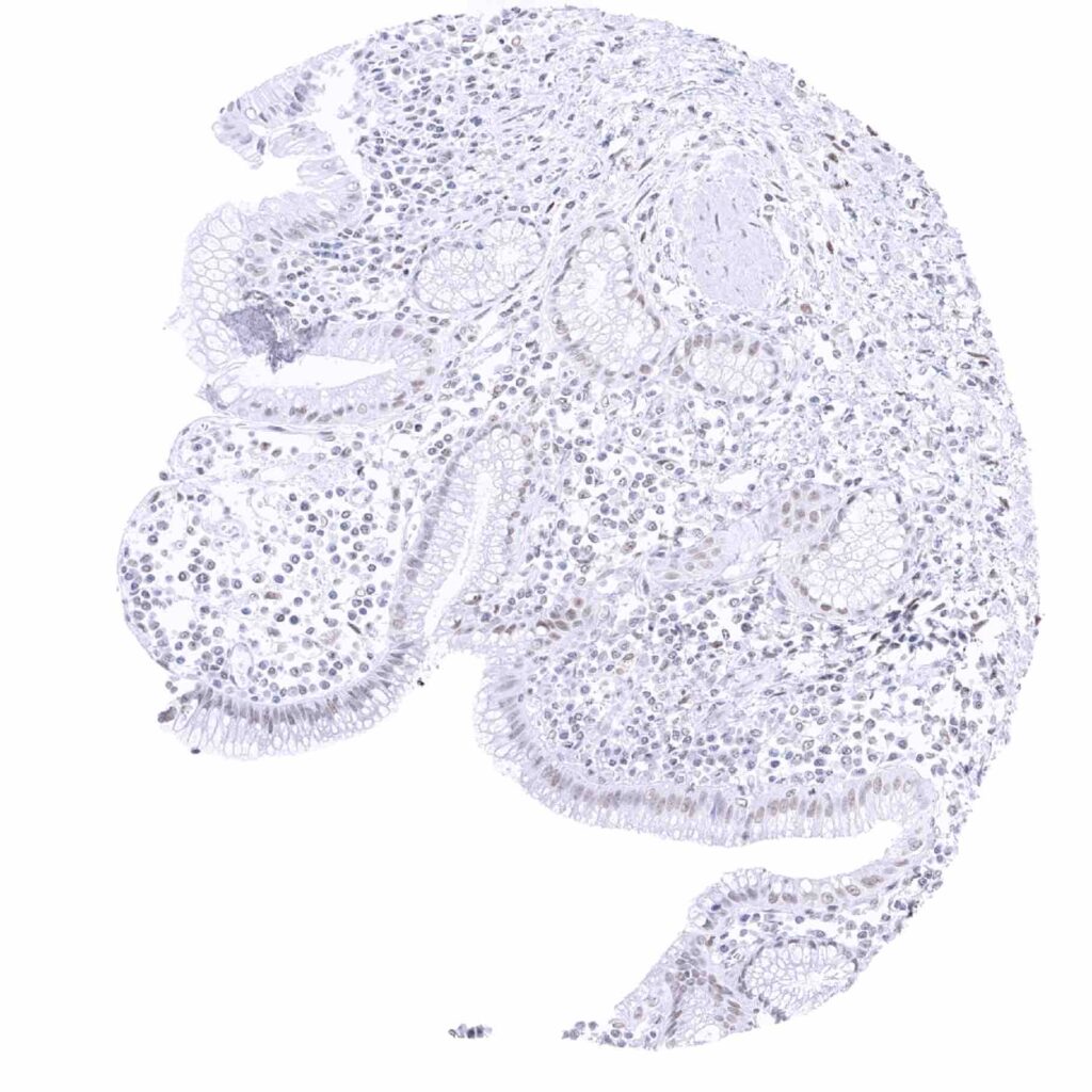
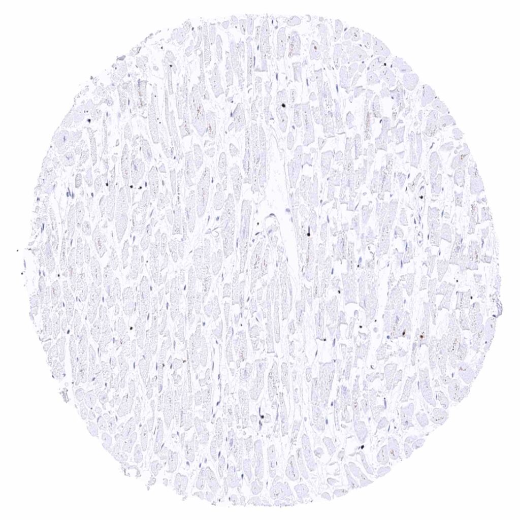
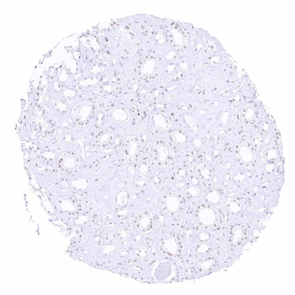
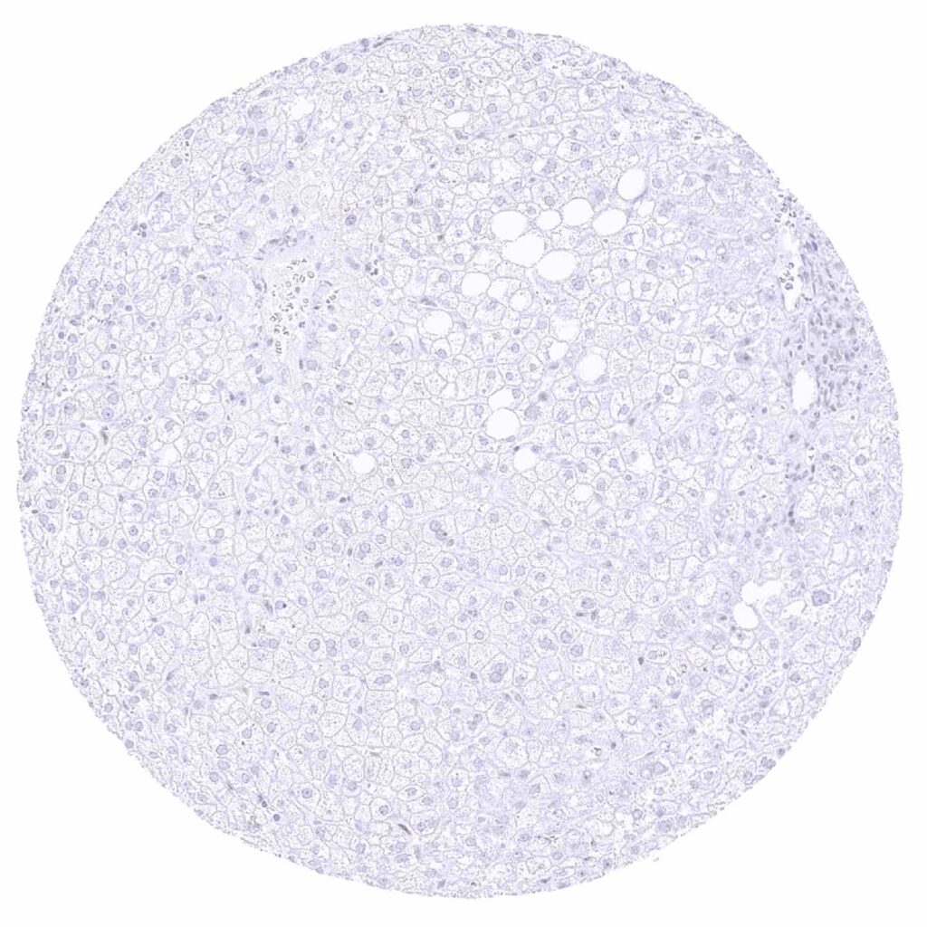
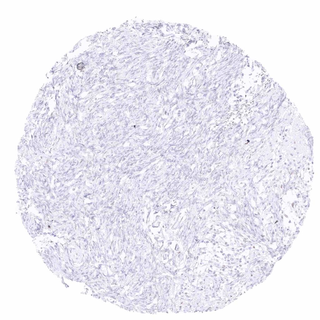
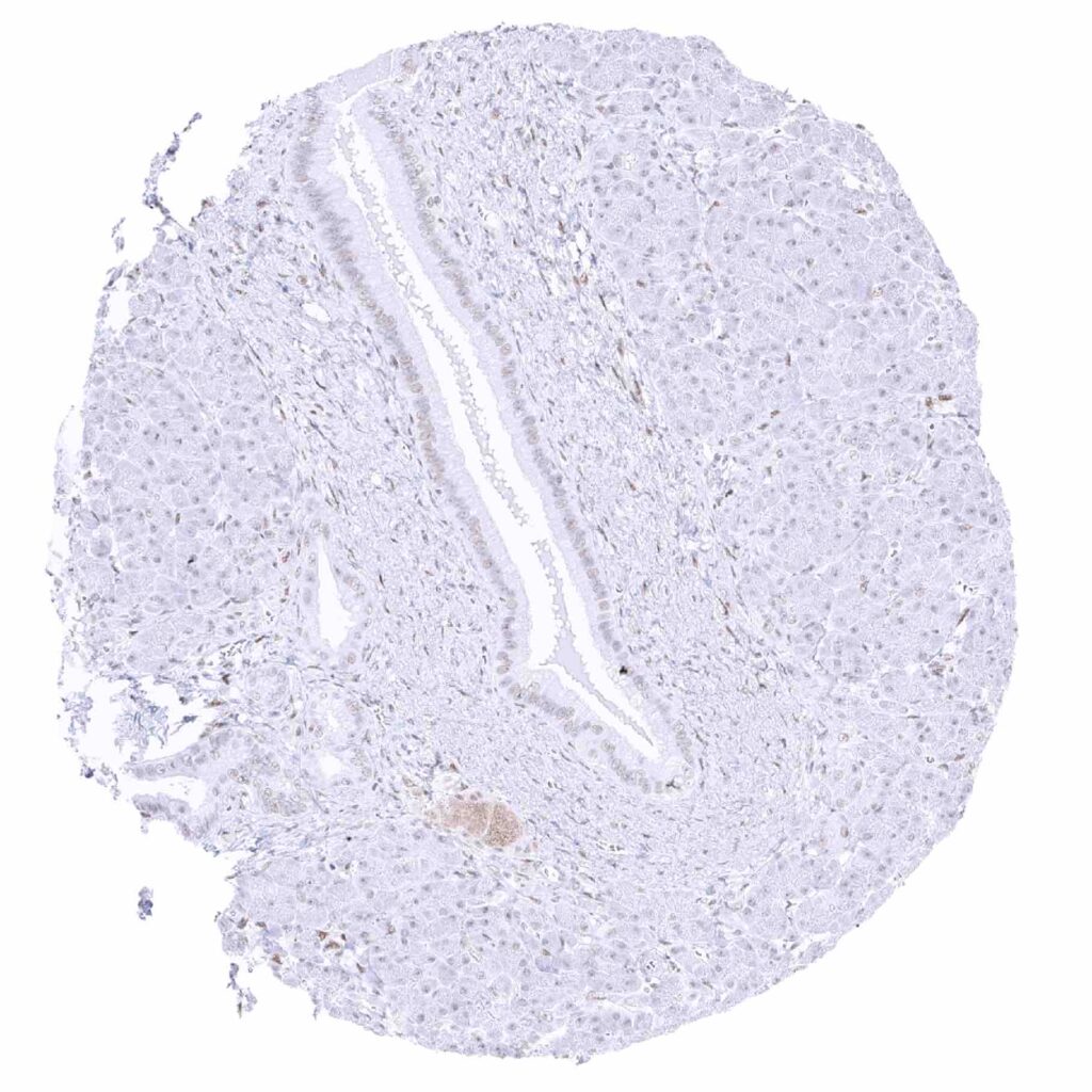
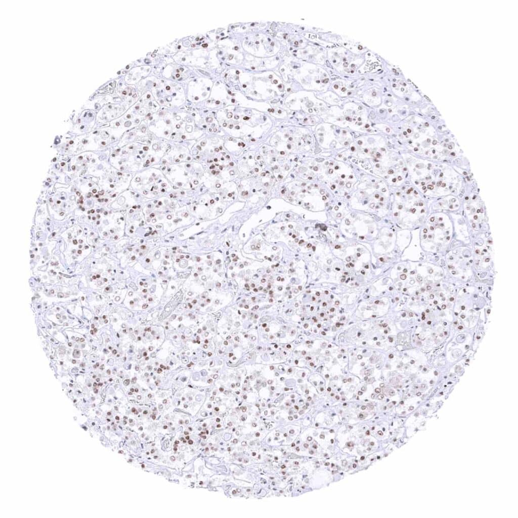
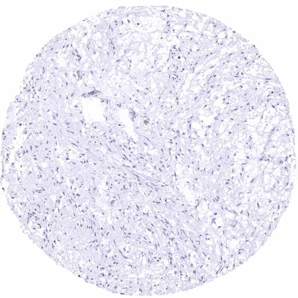
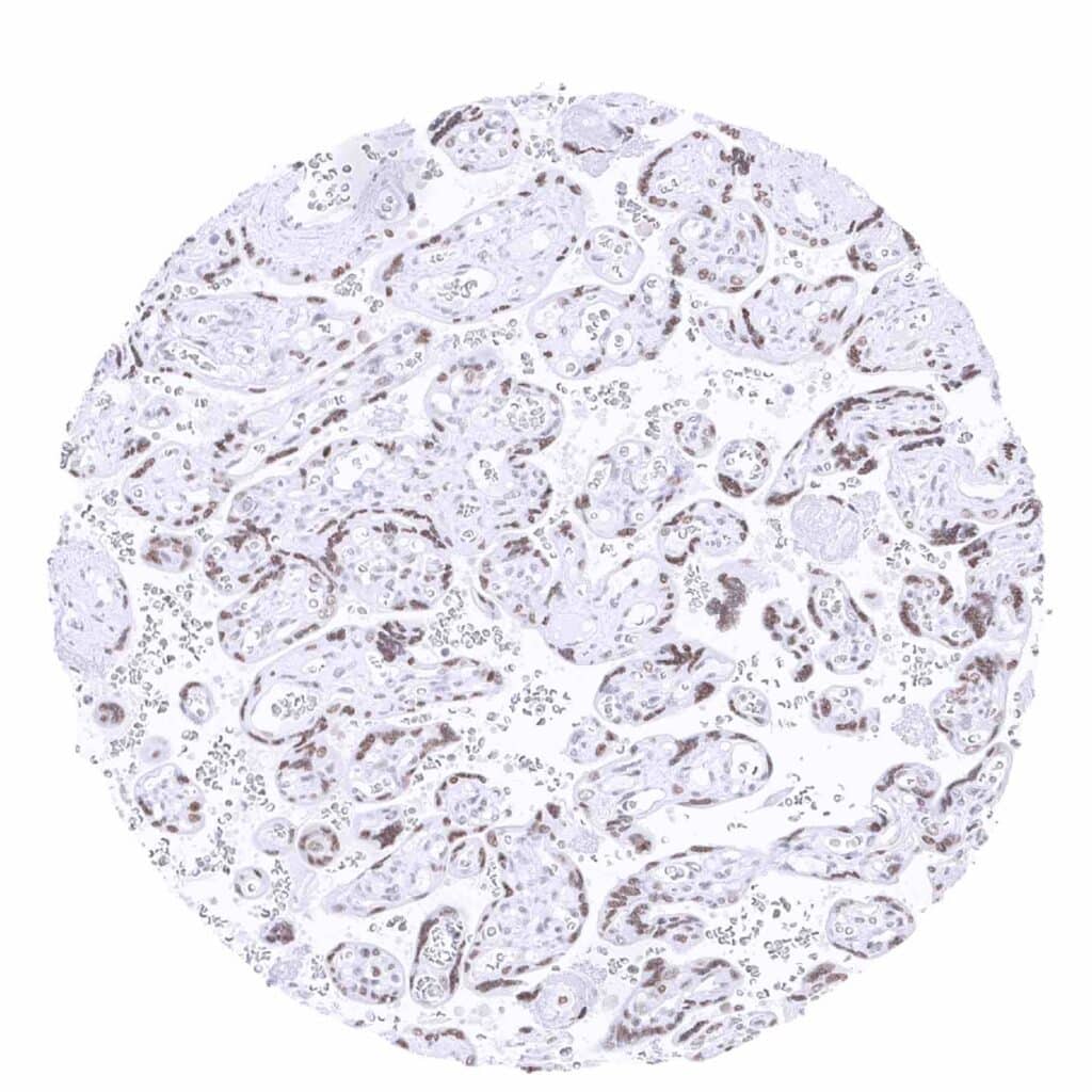
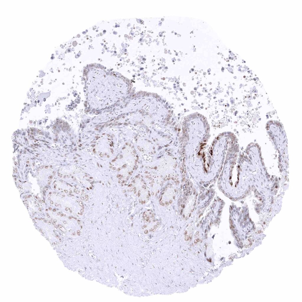
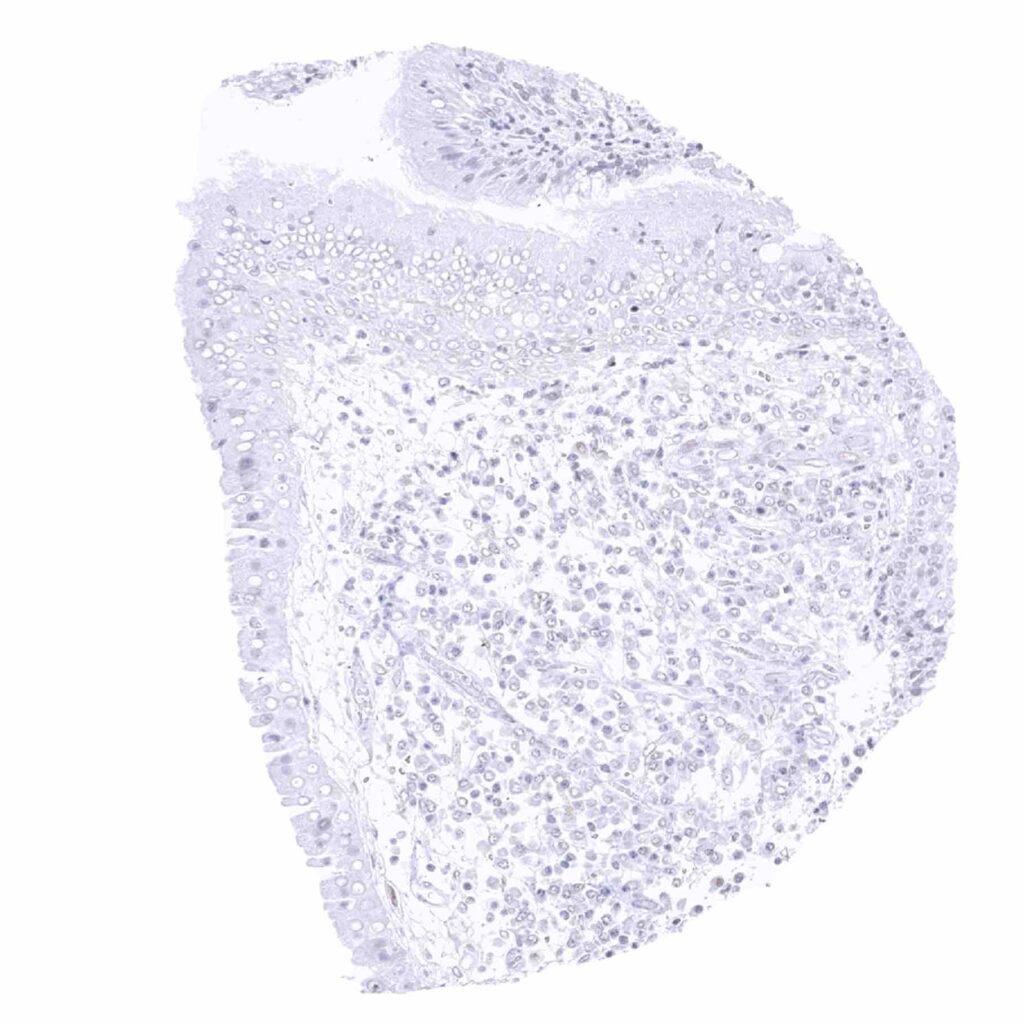
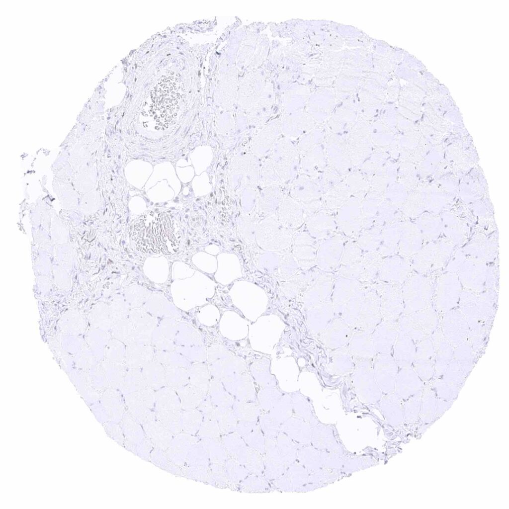
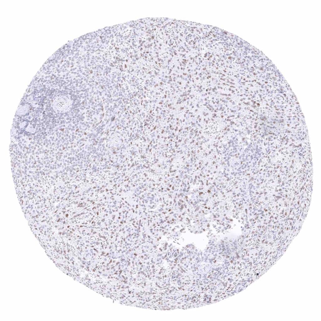
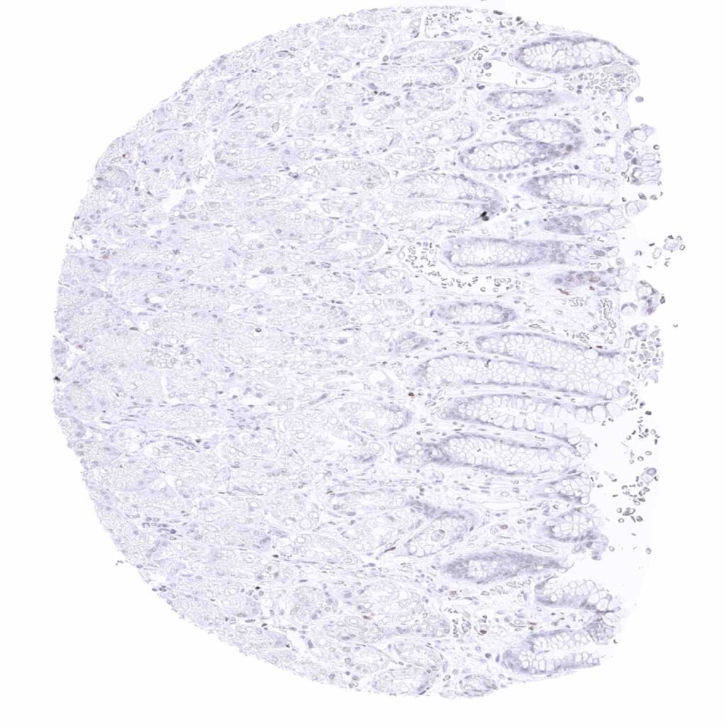
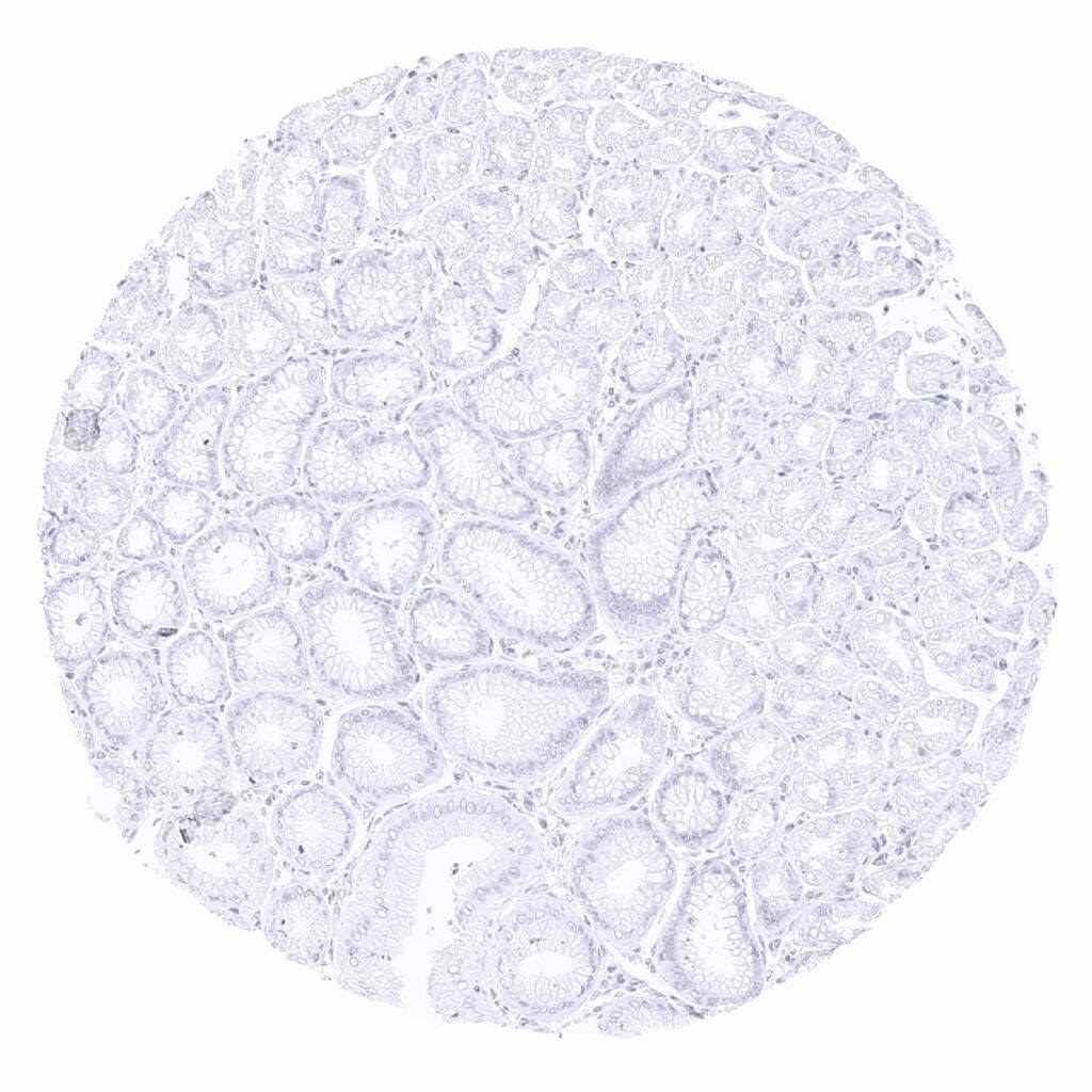
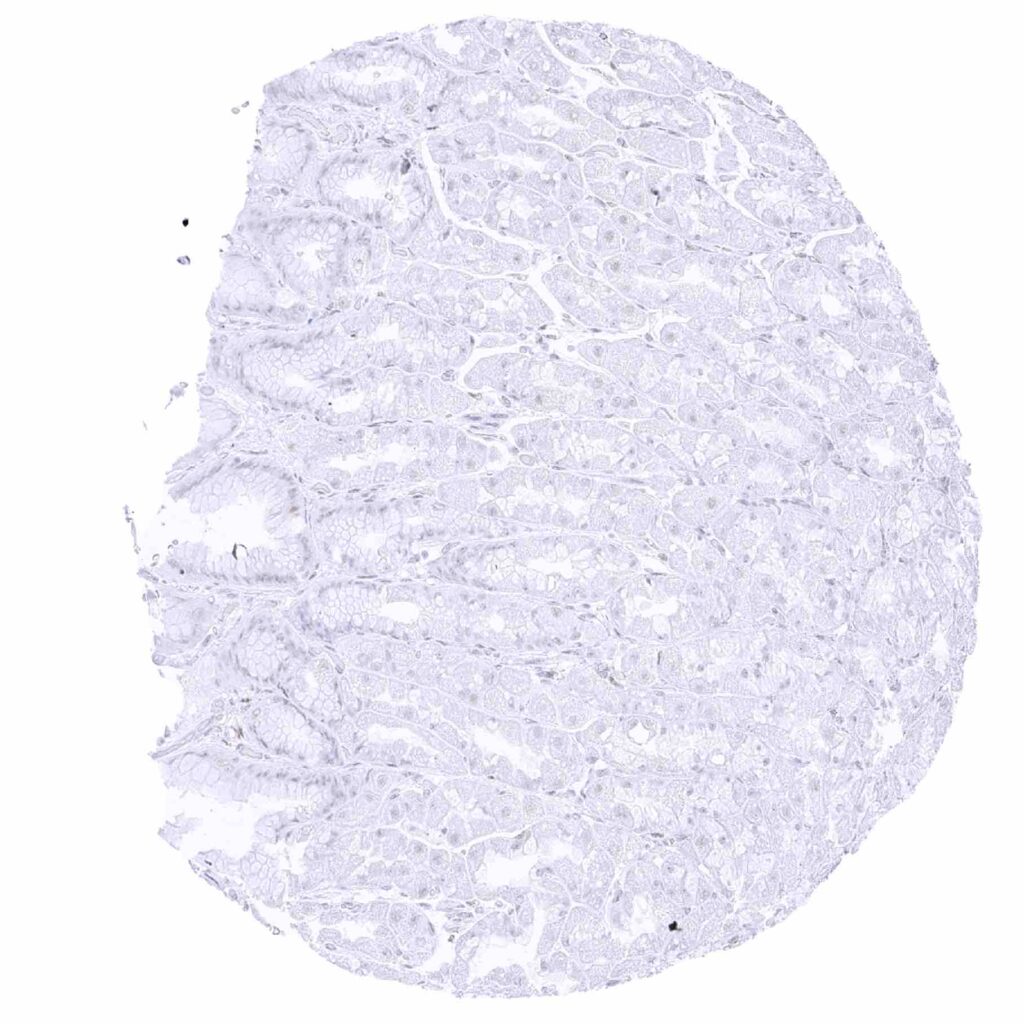
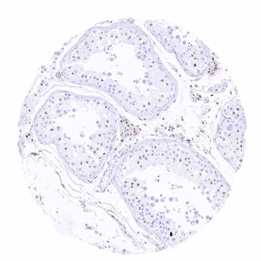
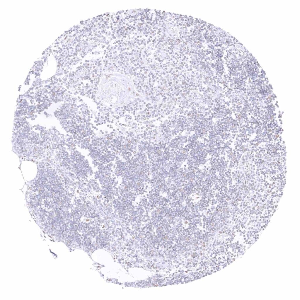
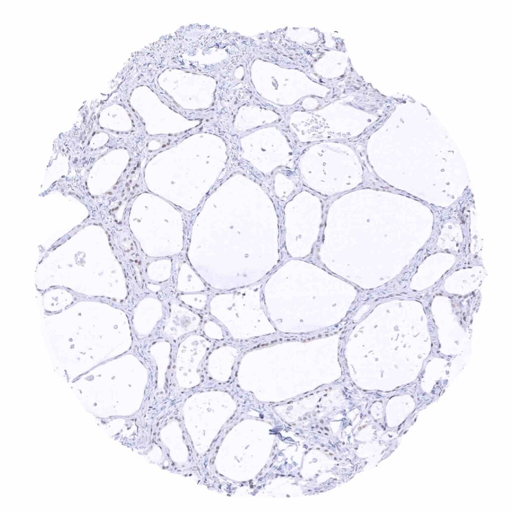
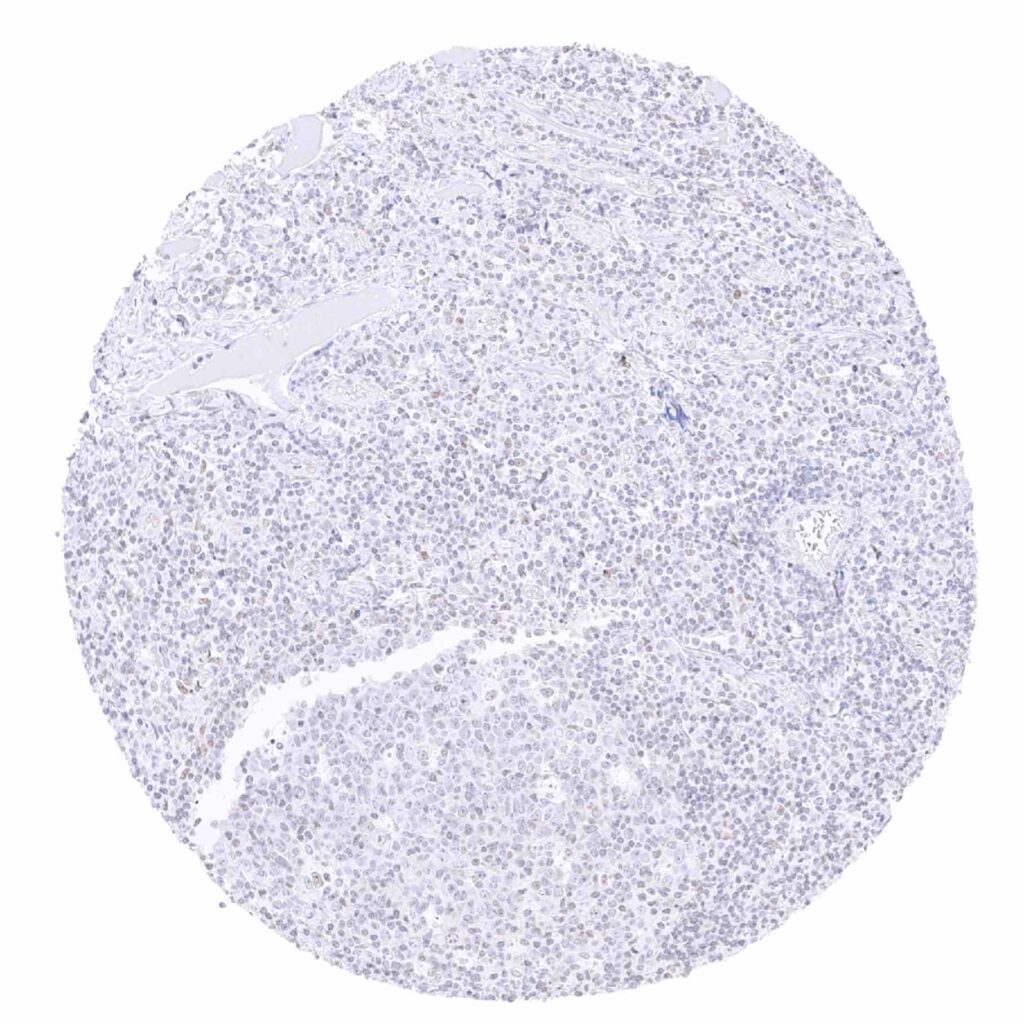
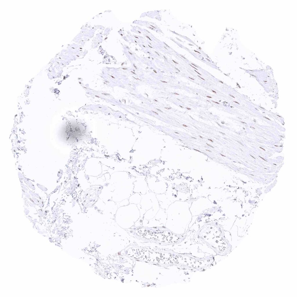
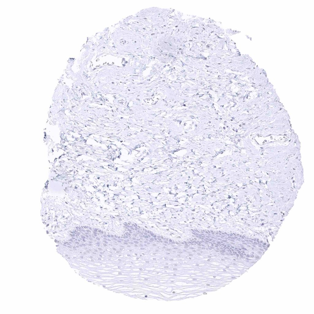
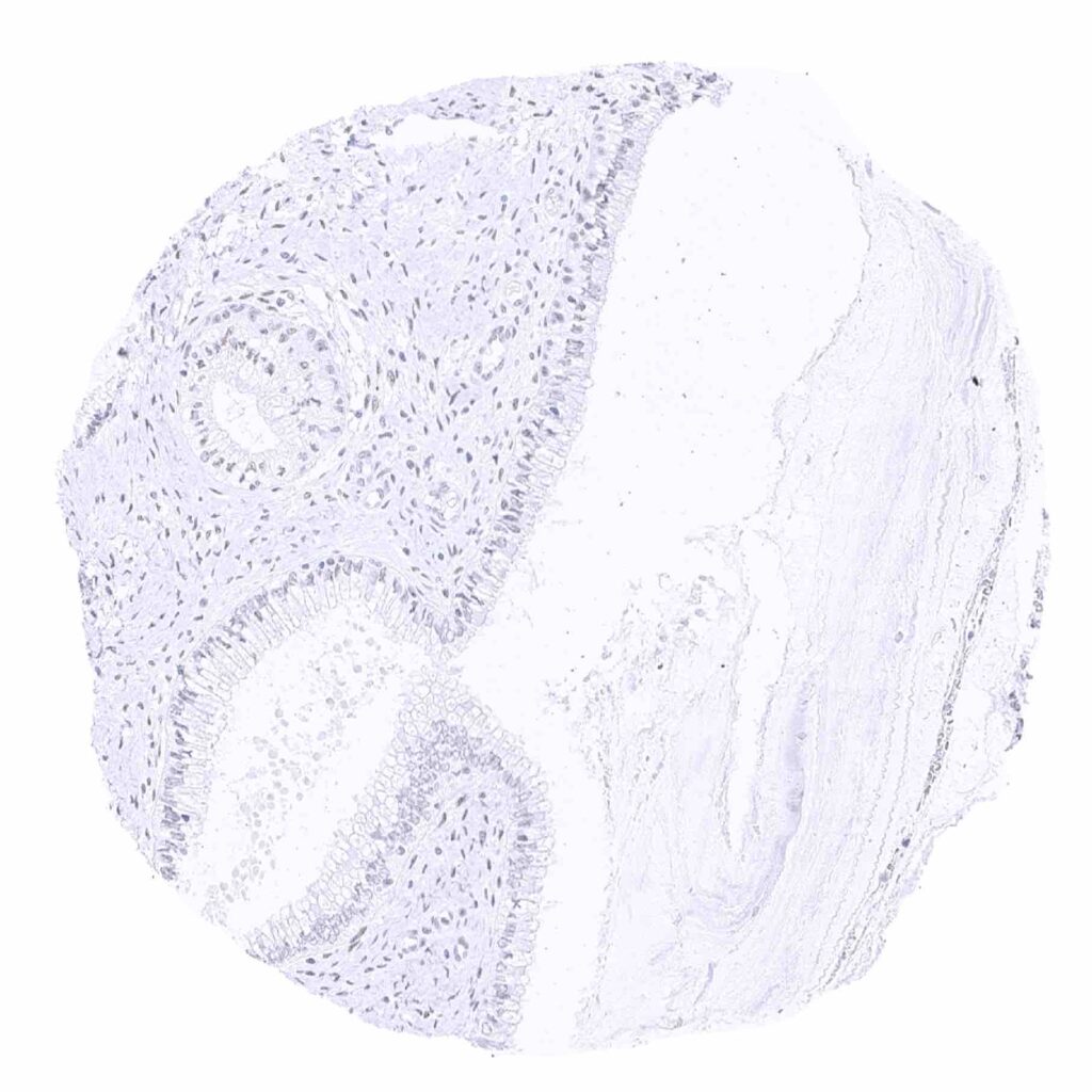
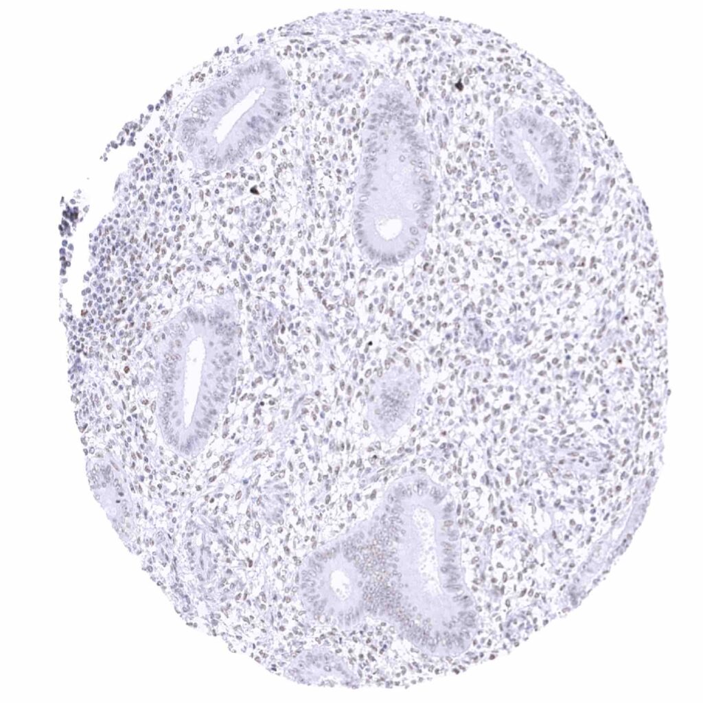
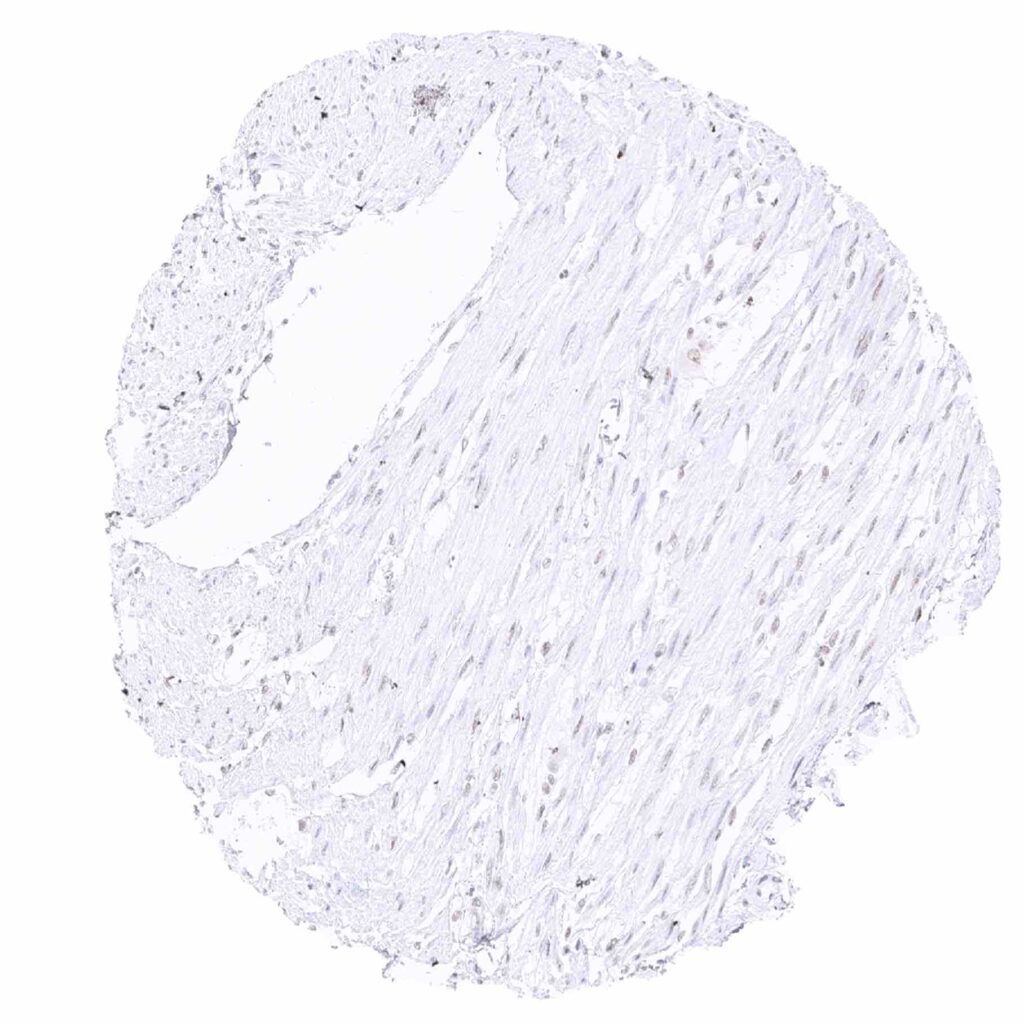
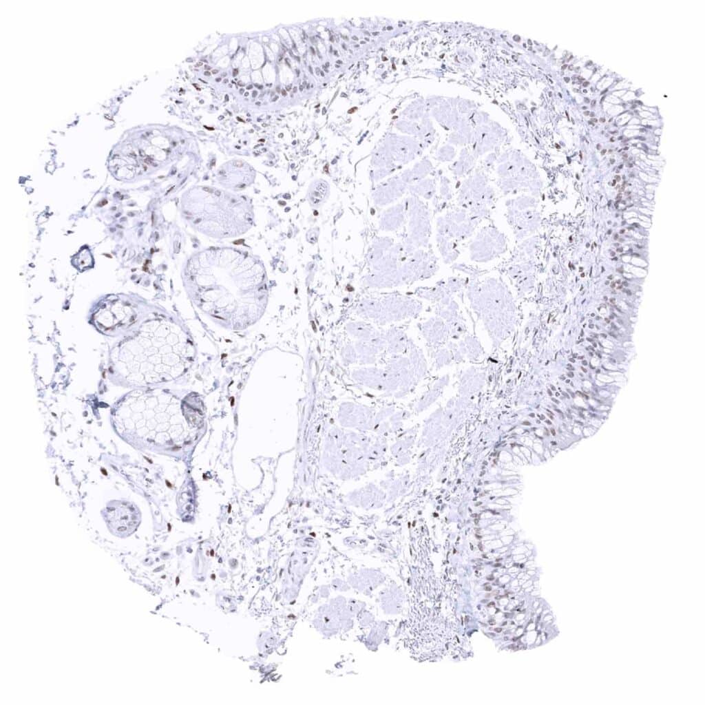
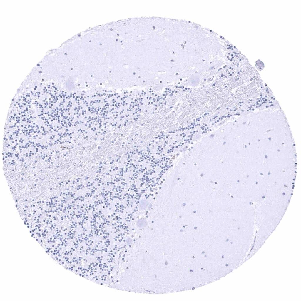
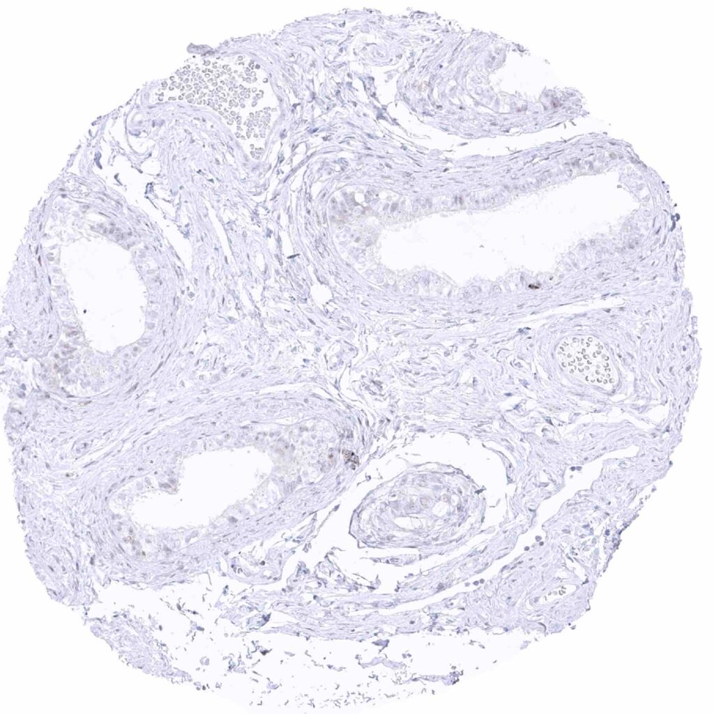
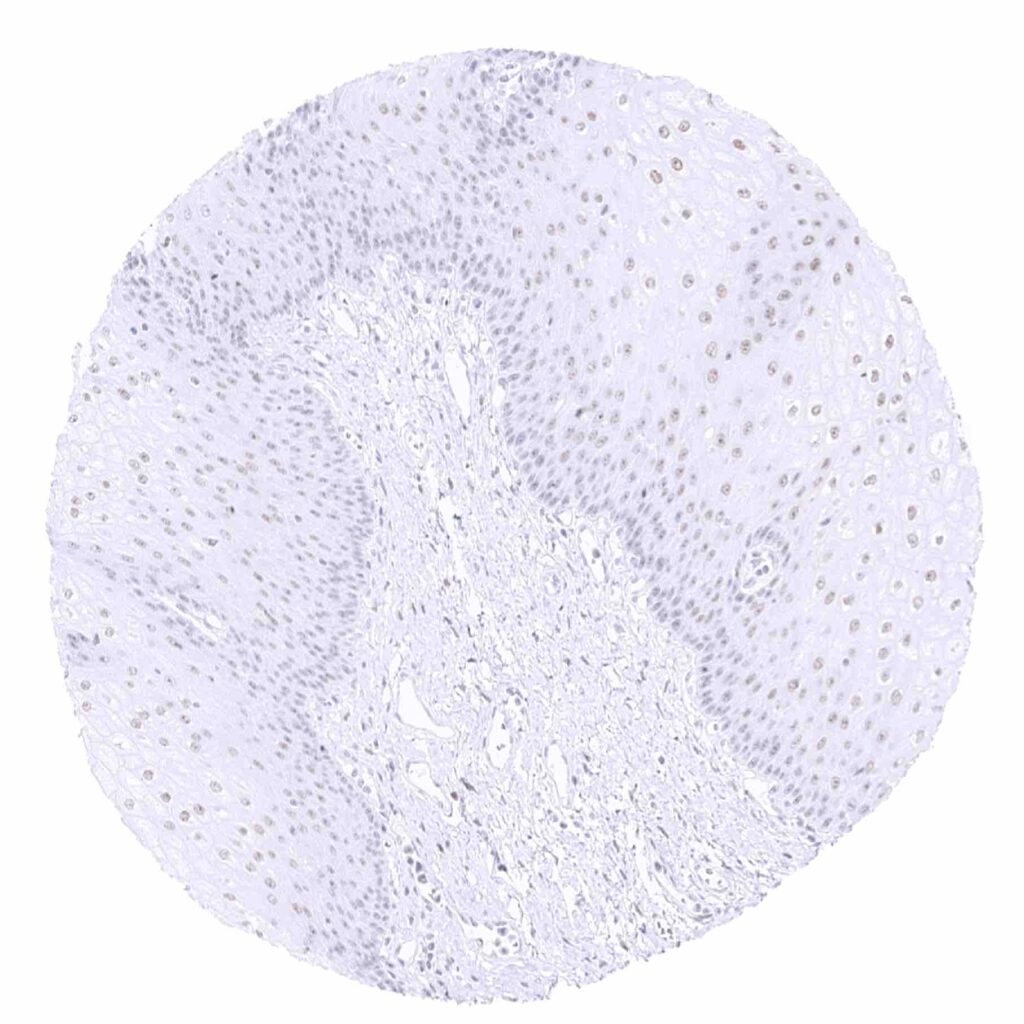
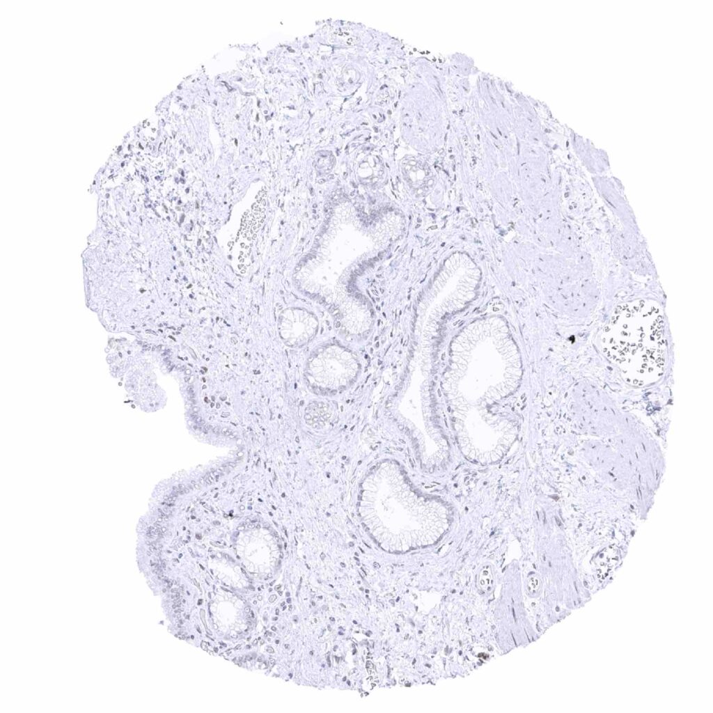
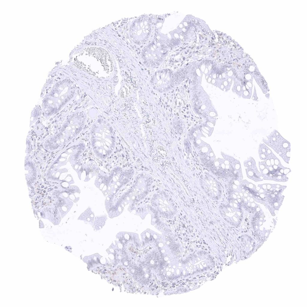
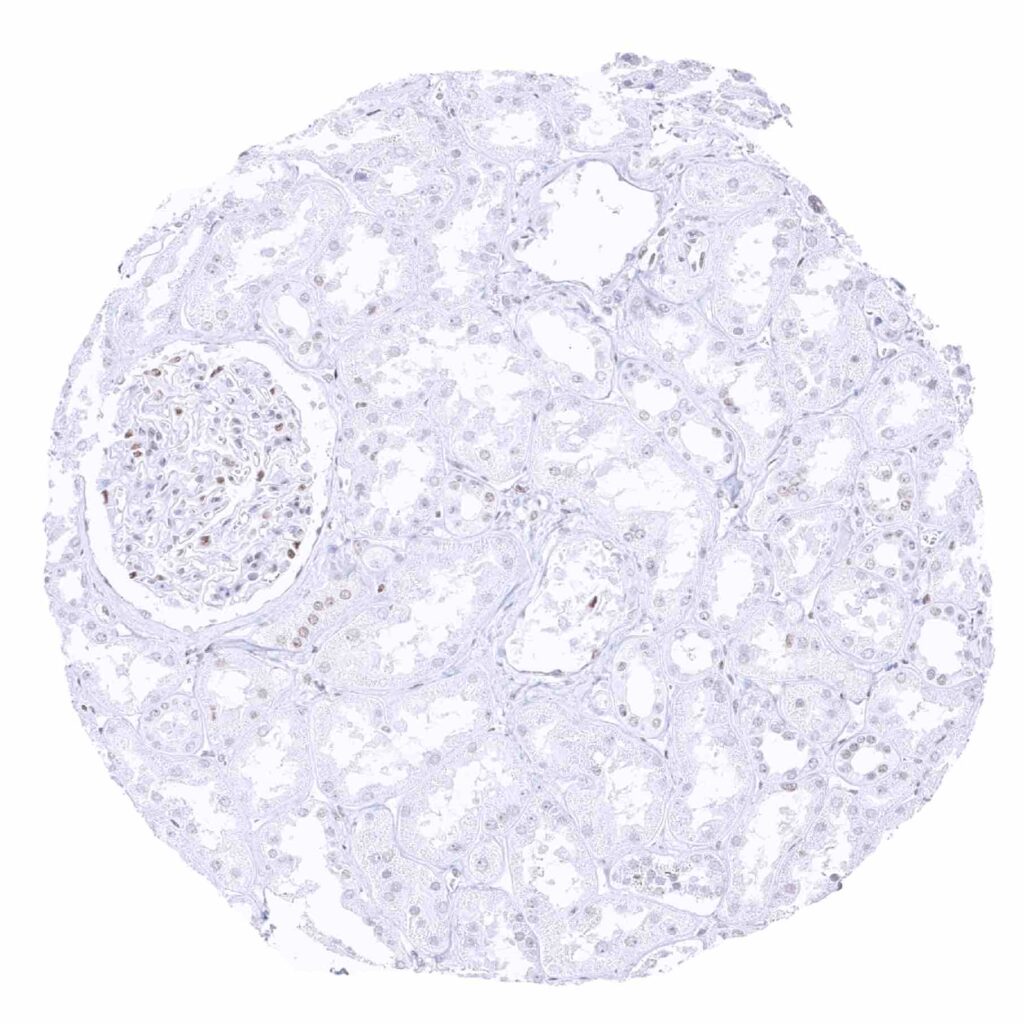
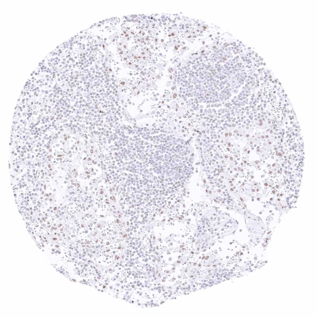
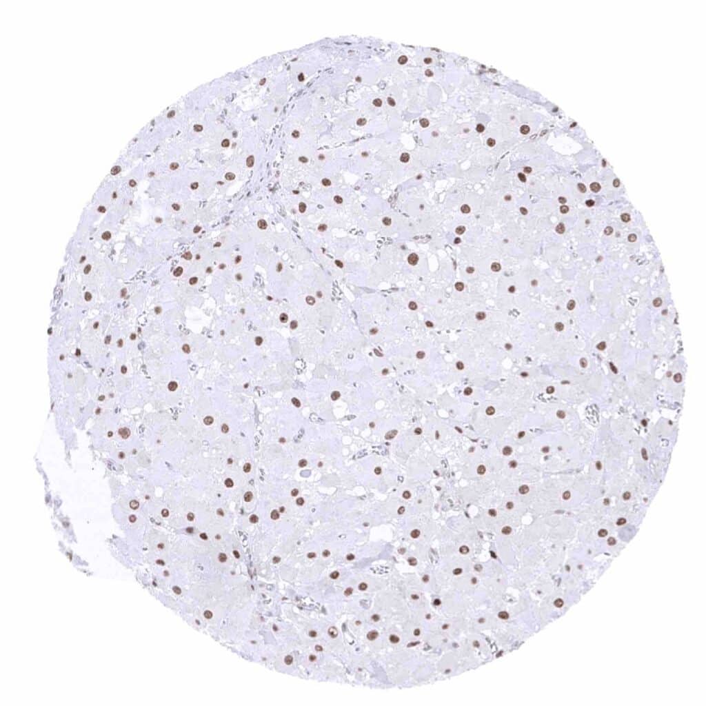
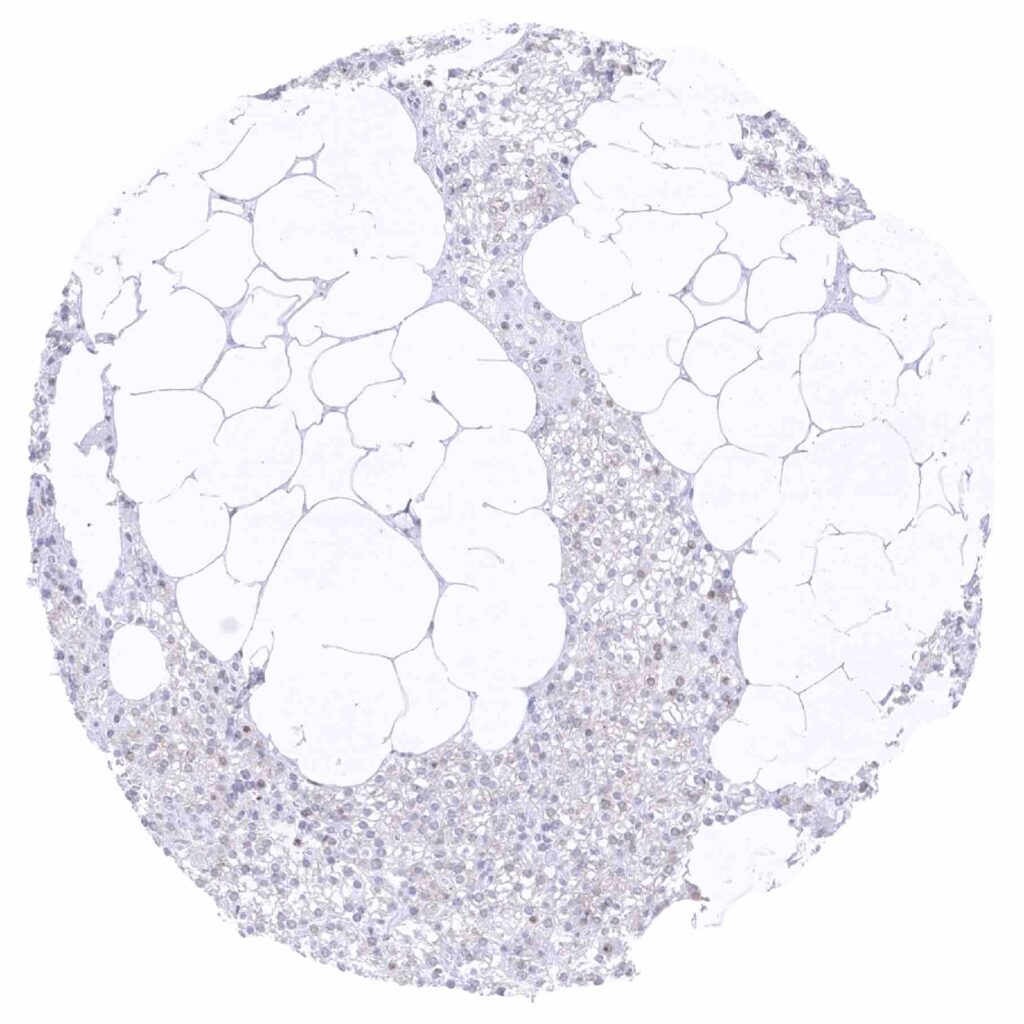
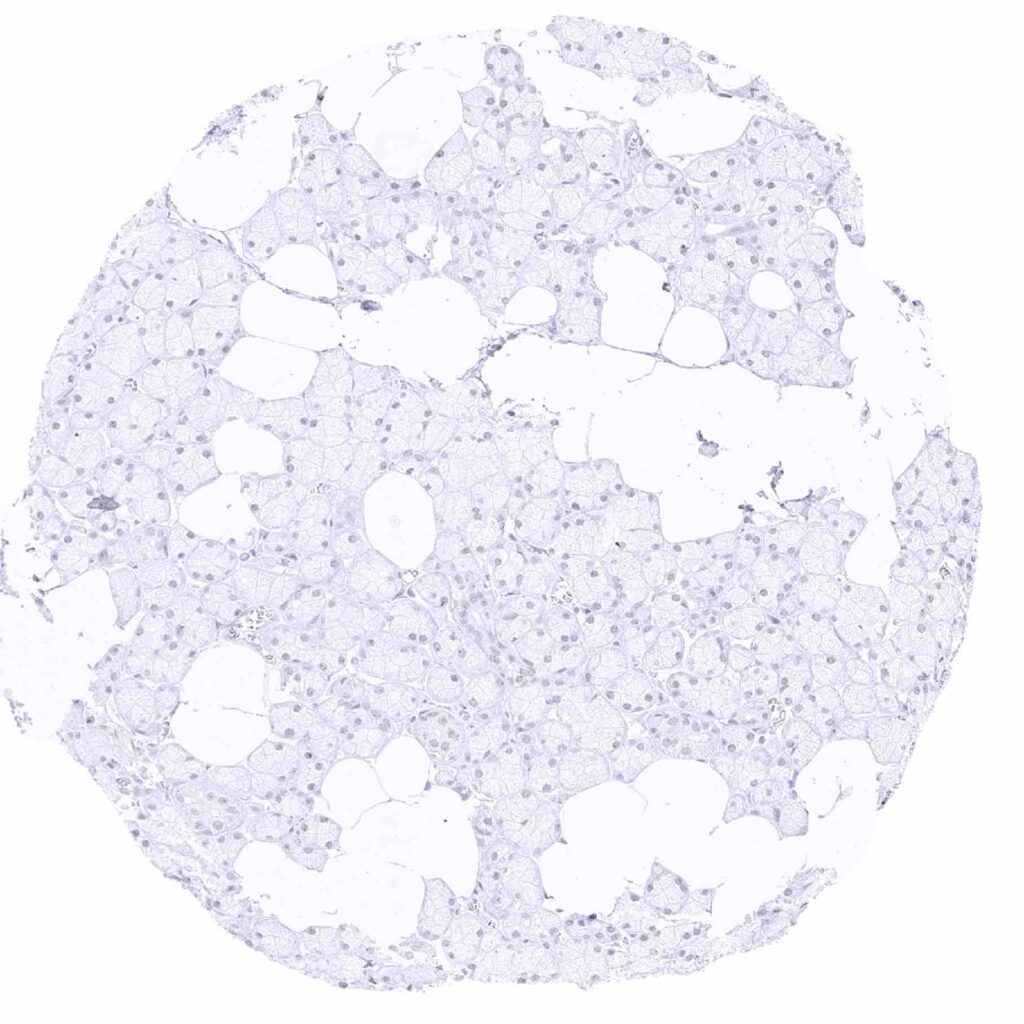
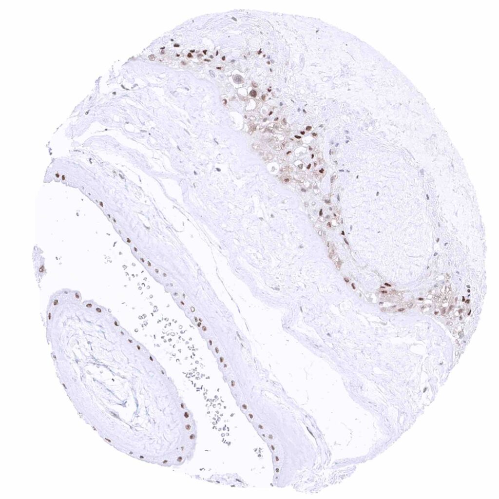
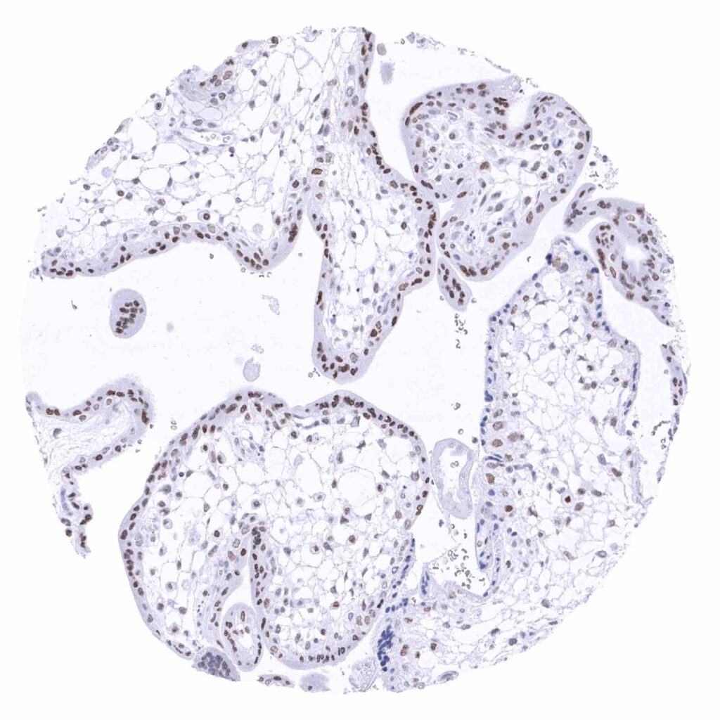
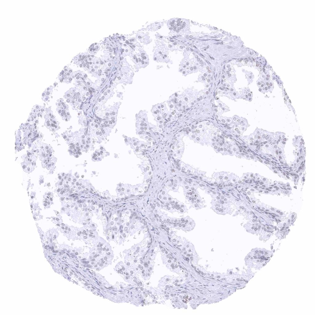
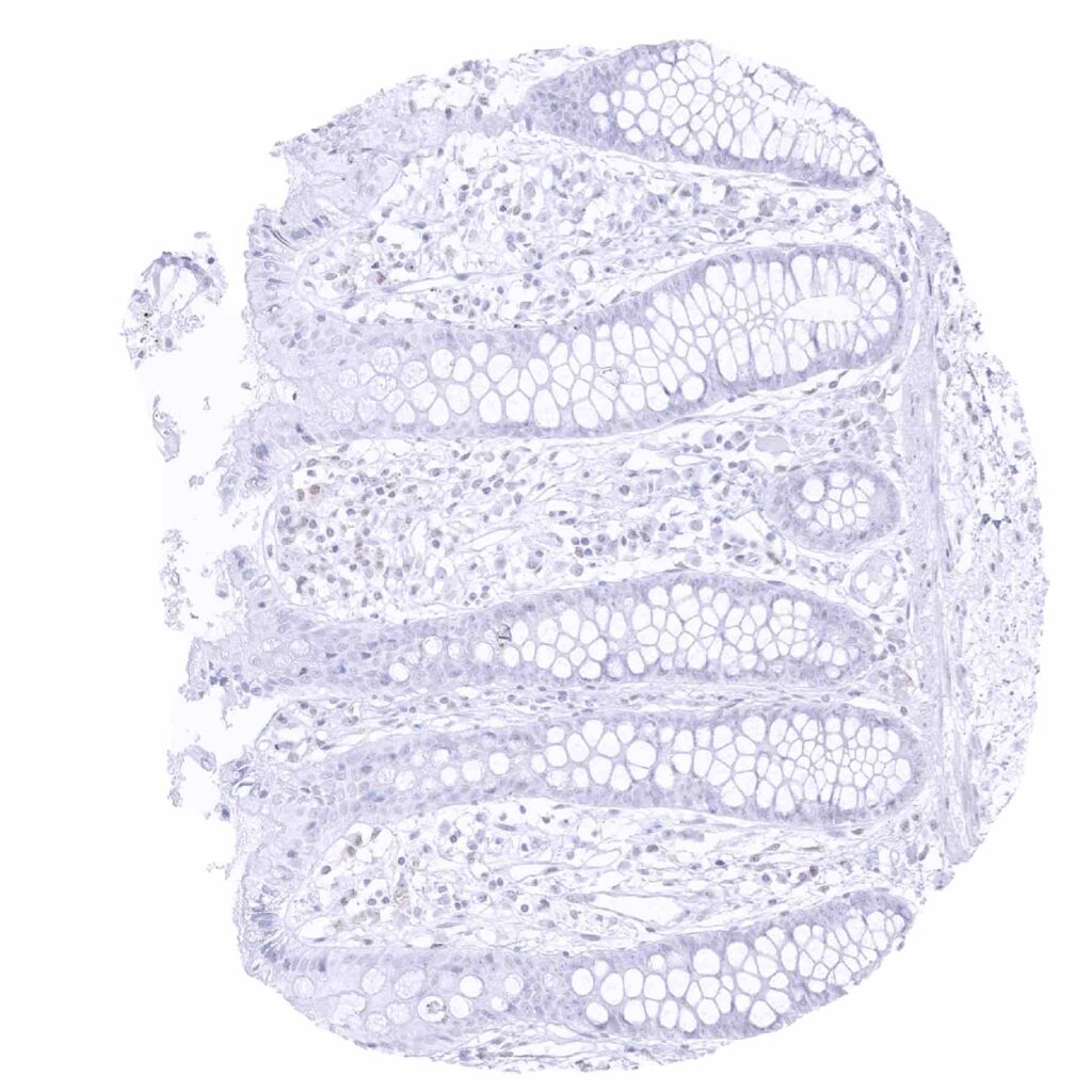
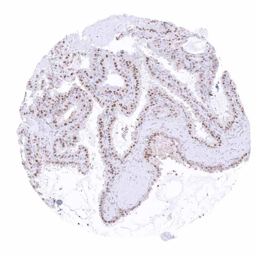
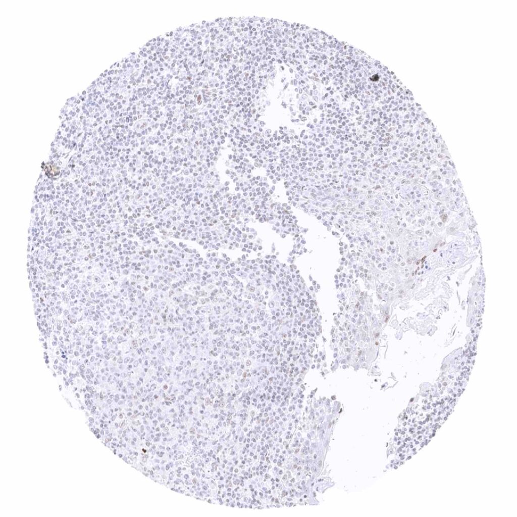
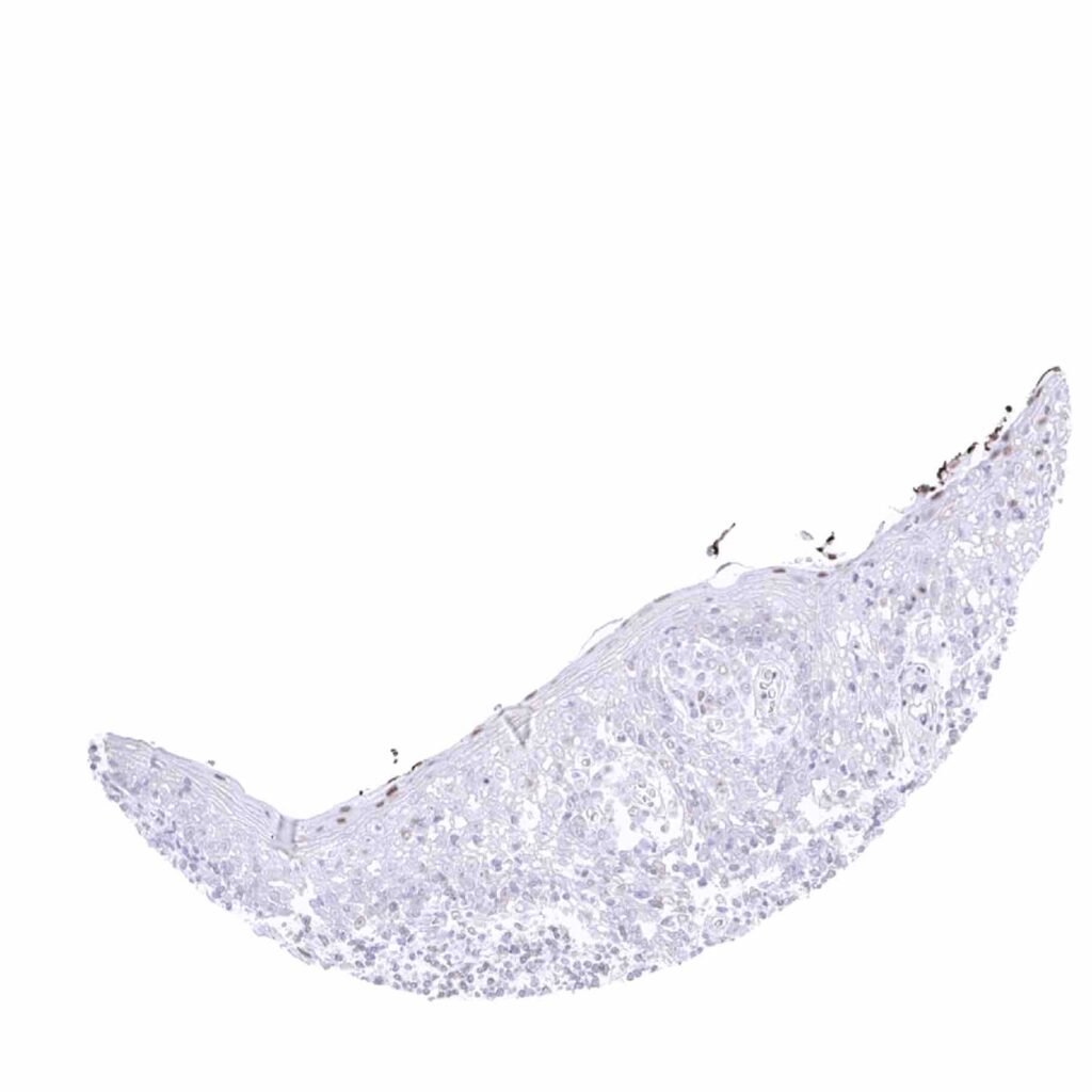
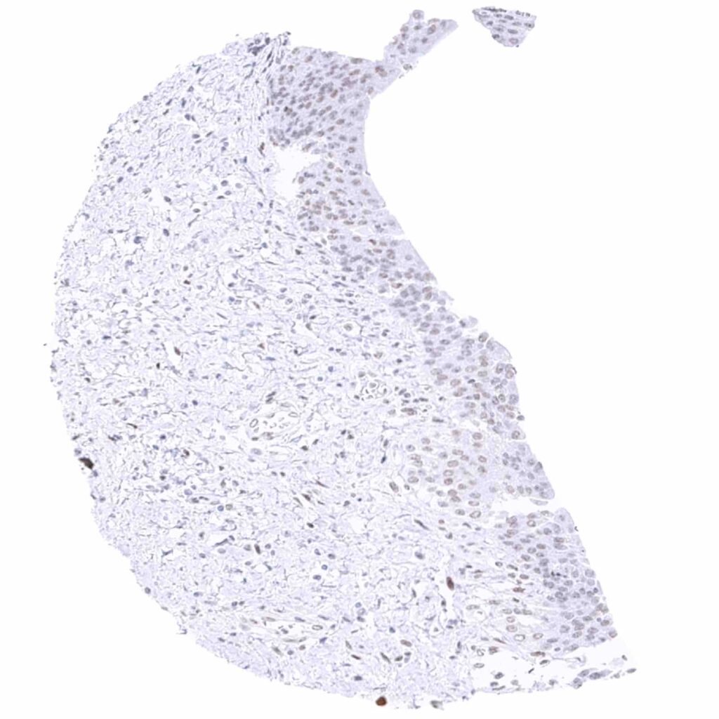
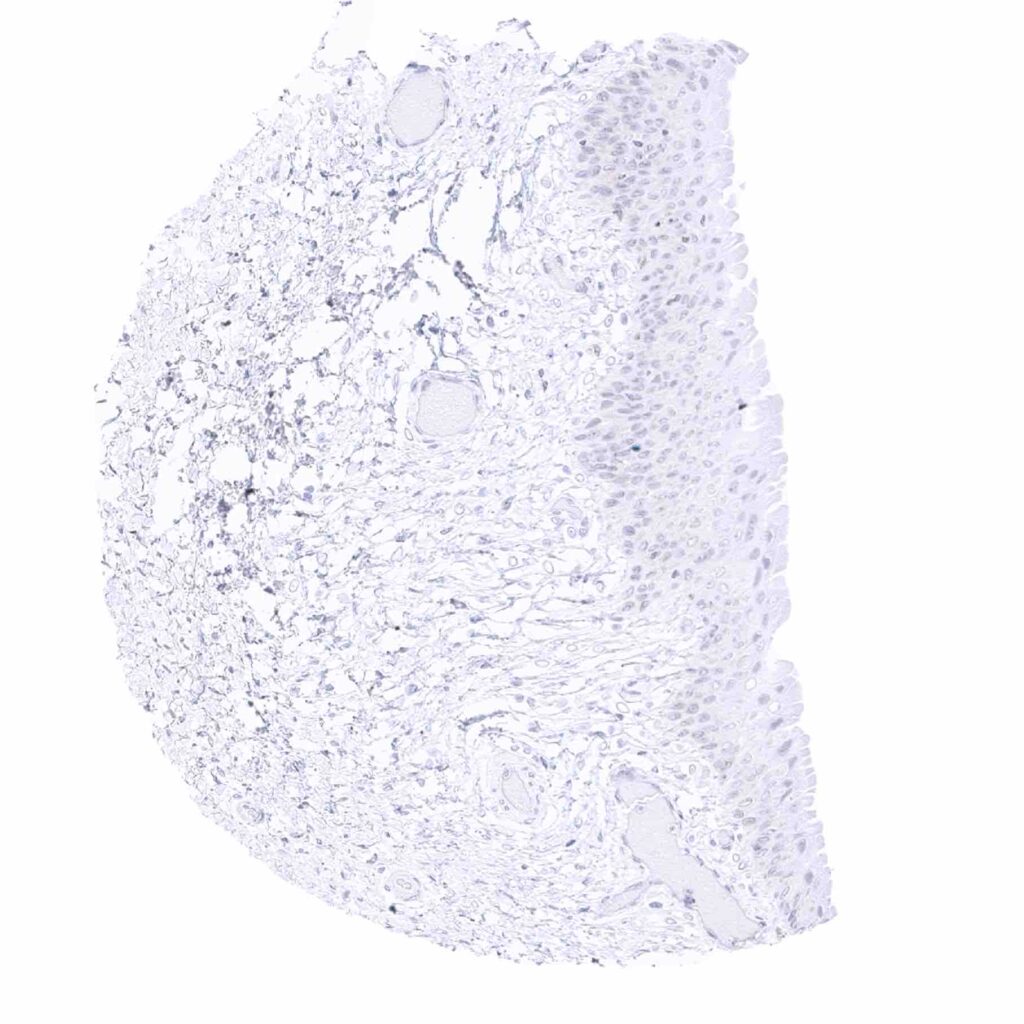
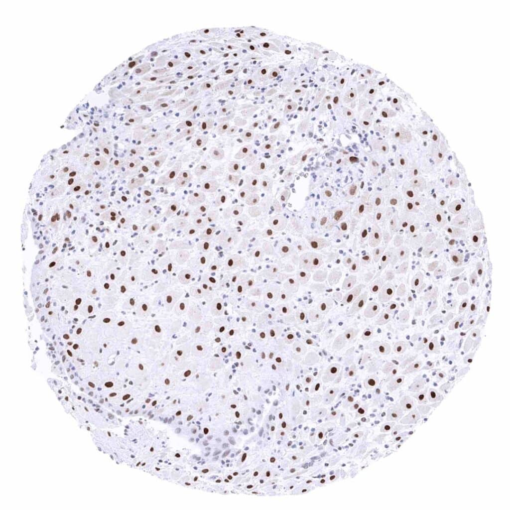
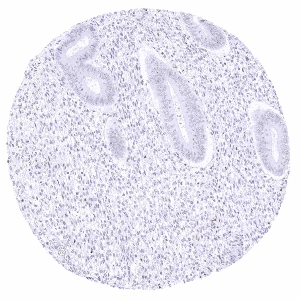
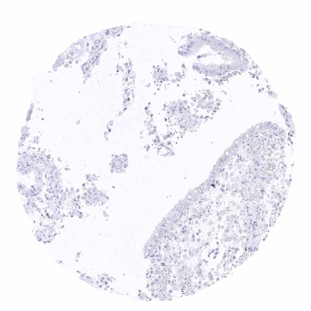
TFE3 antibody [MSVA-403R] HistoMAX™


Go to Cancer Tissue Gallery
  
 Go back to product page
  

Go to HistoMAX page


Adrenal gland – Moderate to strong TFE3 staining of cortical cells

Anal canal, skin

Aorta, media

Appendix, mucosa


Appendix, muscular wall – Faint TFE3 staining of some muscle cells

Bone marrow – Few cells show weak staining

Breast

Bronchus, mucosa – Weak to moderate TFE3 staining of predoinantly basal epithelial cells of the respiratory epithelium and of glandular cells


Cerebellum (molecular layer, Purkinje cell layer, granule cell layer, white matter)

Cerebrum – Some astrocytic cells show faint TFE3 positivity

Colon descendens, mucosa – Some surface epithelial exhibit a faint TFE3 staining while inflammatory cells (macrophages_) show a moderate positivity

Colon descendens, muscular wall


Duodenum, Brunner gland

Duodenum, mucosa

Epididymis – Strong TFE3 staining of epithelial cells in the corpus epididymis

Epididymis – TFE3 staining is absent in the cauda


Esophagus, squamous epithelium – Faint TFE3 staining of most squamous epithelial cells

Fallopian tube, mucosa – Weak nuclear TFE3 staining of a subset of epithelial cells in this sample

Fallopian tube, mucosa

Fat


Gallbladder, epithelium – Absence of TFE3 staining in this sample

Gallbladder, epithelium – Weak TFE3 staining of epithelial cells in a sample showing major inflammation

Heart muscle

Ileum, mucosa


Kidney, cortex – Moderate to strong TFE3 staining of a subset of glomerular cells. Weak TFE3 positivity of a subset of tubular and collecting duct cells

Kidney, medulla – Weak TFE3 positivity of mostcollecting duct cells

Liver

Lung – A large subset of pneumocytes and of alveolar macrophages show weak to moderate TFE3 positivity


Lymph node – Weak to moderateTFE3 staining of a seizable subset of sinusoidal cells (macrophages_)

Ovary, corpus luteum – Intense nuclear TFE3 staining of corpus luteum cells

Ovary, follicular cyst – Strong nuclear TFE3 staining of theca interna cells while the staining is markedly weaker in granulosa cells

Ovary, stroma


Pancreas – Faint nuclear TFE3 staining in acinar and ductal cells, in a sample with inflammation and fibrosis

Parathyroid gland – Some epithelial cells can show a faint TFE3 staining

Parotid gland – Few glandular cells show weak TFE3 staining

Pituitary gland, anterior lobe – Moderate TFE3 staining of the majority of epithelial cells in this sample


Pituitary gland, posterior lobe

Placenta (amnion and chorion) – Moderate to strong TFE3 positivity of chorion and amnion cells

Placenta, early – Strong TFE3 staining of syncytiotrophoblast cells while TFE3 positivity is markedly weaker for cytotrophoblast and stroma cells

Placenta, mature – Strong TFE3 staining of trophoblast cells


Prostate

Rectum, mucosa

Seminal vesicle – Moderate to strong TFE3 staining of epithelial cells

Seminal vesicle – Strong TFE3 staining of only some groups of epithelial cells in this sample


Sinus paranasales – Absence of TFE3 staining in this sample

Skeletal muscle – Very faint TFE3 staining of some muscle cells

Spleen – Weak to moderate TFE3 staining of sinusoidal cells (macrophages_)

Stomach, antrum


Stomach, antrum

Stomach, corpus – Faint TFE3 staining of mucous neck cells

Testis – Moderate TFE3 staining of Sertoli and Leydig cells

Thymus – Weak TFE3 staining of few squamous epithelial cells in corpuscles of Hassall's


Thyroid gland – Weak to moderate TFE3 staining of most follicular cells

Tonsil – Weak TFE3 staining of the most superficial cell layers of the squamous epithelium

Tonsil, surface epithelium – Moderate TFE3 staining of few superficial epithelial cells

Tonsil


Urinary bladder, muscular wall – Weak TFE3 staining of smooth muscle cells

Urinary bladder, urothelium – Faint TFE3 positivity of some urothelial cells

Urinary bladder, urothelium

Uterus, ectocervix


Uterus, endocervix

Uterus, endometrium (pregnancy) – Intense nuclear TFE3 positivity in decidua cells

Uterus, endometrium (proliferation)

Uterus, endometrium (proliferation)


Uterus, endometrium (secretion)

Uterus, myometrium – Faint TFE3 staining of some muscle cells


| TOP |