GeneTex
  • Country / Location Selection

United States (US)

PD-L1 antibody

Anti-PD-L1 antibody used in Western Blot (WB). GTX104763
Anti-PD-L1 antibody used in Immunocytochemistry/ Immunofluorescence (ICC/IF). GTX104763
Anti-PD-L1 antibody used in Western Blot (WB). GTX104763
Anti-PD-L1 antibody used in IHC (Paraffin sections) (IHC-P). GTX104763
Anti-PD-L1 antibody used in Immunocytochemistry/ Immunofluorescence (ICC/IF). GTX104763
Anti-PD-L1 antibody used in IHC (Paraffin sections) (IHC-P). GTX104763
Anti-PD-L1 antibody used in Western Blot (WB). GTX104763
Anti-PD-L1 antibody used in IHC (Paraffin sections) (IHC-P). GTX104763
Anti-PD-L1 antibody used in Western Blot (WB). GTX104763

Cat. No. GTX104763

Host

Rabbit

Clonality

Polyclonal

Isotype

IgG

Application

WB, ICC/IF, IHC-P, IHC-Fr, FACS

Reactivity

Human
Package
100 μl ($399),
25 μl ($169)
PRODUCT

Summary

PD-L1 antibody recognizes programmed death-ligand 1 (PD-L1) protein, also known as B7 homolog 1 (B7-H1) and cluster of differentiation 274 (CD274) protein. PD-L1 is found on various cancer cells and transduces immunosuppressive signals by binding to the programmed cell death protein 1 (PD-1) on effector T cells, thereby diminishing immune system attacks on malignant cells. Blocking the interaction between PD-L1 and PD-1 with PD-L1 antibodies is generating great clinical interest as a mode of immunotherapy against a number of cancers.

APPLICATION

Application Note

*Optimal dilutions/concentrations should be determined by the researcher.
Application Recommended Dilution
WB 1:500-1:3000
ICC/IF 1:100-1:1000
IHC-P 1:100-1:1000
IHC-Fr Assay dependent
FACS Assay dependent
Not tested in other applications.

Calculated MW

33 kDa. ( Note )

Observed MW

50 kDa. The observed M.W. is based on the following publication. PMID: 23674495

Positive Control

MDA-MB-231 (24μg/ml Tunicamycin treatment for 16 hr) , MDA-MB-231 , A431(100 ng/mL IFN-gamma treatment for 48 hr)

Product Note

This antibody is specific for human PDL1 protein, and it does not cross react with human PDL2 protein.

PROPERTIES

Form

Liquid

Buffer

PBS, 20% Glycerol

Preservative

0.025% ProClin 300

Storage

Store as concentrated solution. Centrifuge briefly prior to opening vial. For short-term storage (1-2 weeks), store at 4ºC. For long-term storage, aliquot and store at -20ºC or below. Avoid multiple freeze-thaw cycles.

Concentration

0.97 mg/ml (Please refer to the vial label for the specific concentration.)

Antigen Species

Human

Immunogen

Synthetic peptide encompassing a sequence within the Intracellular domain of human PD-L1. The exact sequence is proprietary.

Purification

Purified by antigen-affinity chromatography.

Conjugation

Unconjugated

RRID

AB_1240586

Note

For laboratory research use only. Not for any clinical, therapeutic, or diagnostic use in humans or animals. Not for animal or human consumption.

Purchasers shall not, and agree not to enable third parties to, analyze, copy, reverse engineer or otherwise attempt to determine the structure or sequence of the product.

TARGET

Synonyms

CD274 molecule , B7-H , B7H1 , PD-L1 , PDCD1L1 , PDCD1LG1 , PDL1

Cellular Localization

Isoform 1: Cell membrane; Single-pass type I membrane protein , Isoform 2: Endomembrane system; Single-pass type I membrane protein

Background

Involved in the costimulatory signal, essential for T-cell proliferation and production of IL10 and IFNG, in an IL2-dependent and a PDCD1-independent manner. Interaction with PDCD1 inhibits T-cell proliferation and cytokine production.

Database

Research Area

DATA IMAGES

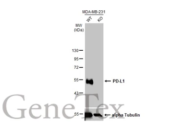
Anti-PD-L1 antibody used in Western Blot (WB). GTX104763

GTX104763 WB Image

Wild-type (WT) and PD-L1 knockout (KO) MDA-MB-231 cell extracts (30 μg) were separated by 10% SDS-PAGE, and the membrane was blotted with PD-L1 antibody (GTX104763) diluted at 1:4000. The HRP-conjugated anti-rabbit IgG antibody (GTX213110-01) was used to detect the primary antibody.

Anti-PD-L1 antibody used in Western Blot (WB). GTX104763

GTX104763 WB Image

Untreated (–) and treated (+) A431 whole cell extracts (30 μg) were separated by 10% SDS-PAGE, and the membranes were blotted with PD-L1 antibody (GTX104763) diluted at 1:1200 and competitor's antibody (CST#13684) diluted at 1:500. The HRP-conjugated anti-rabbit IgG antibody (GTX213110-01) was used to detect the primary antibody.

*The competitor is not affiliated with GeneTex and does not endorse this product.

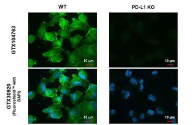
Anti-PD-L1 antibody used in Immunocytochemistry/ Immunofluorescence (ICC/IF). GTX104763

GTX104763 ICC/IF Image

PD-L1 antibody detects PD-L1 protein at cell membrane by immunofluorescent analysis.
Sample: MDA-MB-231 cells were fixed in ice-cold MeOH for 5 min.
Green: PD-L1 stained by PD-L1 antibody (GTX104763) diluted at 1:500.
Blue: Fluoroshield with DAPI (GTX30920).

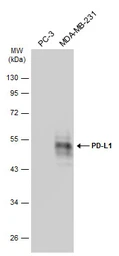
Anti-PD-L1 antibody used in Western Blot (WB). GTX104763

GTX104763 WB Image

Various whole cell extracts (30 μg) were separated by 10% SDS-PAGE, and the membrane was blotted with PD-L1 antibody (GTX104763) diluted at 1:2000. The HRP-conjugated anti-rabbit IgG antibody (GTX213110-01) was used to detect the primary antibody, and the signal was developed with Trident ECL plus-Enhanced.

Anti-PD-L1 antibody used in Western Blot (WB). GTX104763

GTX104763 WB Image

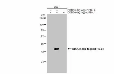
Non-transfected (–) and transfected (+) A431 whole cell extracts (30 μg) were separated by 10% SDS-PAGE, and the membrane was blotted with PD-L1 antibody (GTX104763) diluted at 1:1000. The HRP-conjugated anti-rabbit IgG antibody (GTX213110-01) was used to detect the primary antibody.

Anti-PD-L1 antibody used in Western Blot (WB). GTX104763

GTX104763 WB Image

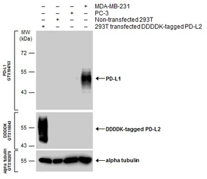
Various whole cell extracts were separated by 10% SDS-PAGE, and the membranes were blotted with PD-L1 antibody (GTX104763) diluted at 1:600 and with DDDDK tag antibody (GTX115043) diluted at 1:3000 to detect DDDDK-tagged PD-L2. The HRP-conjugated anti-rabbit IgG antibody (GTX213110-01) was used to detect the primary antibody.

Anti-PD-L1 antibody used in IHC (Paraffin sections) (IHC-P). GTX104763

GTX104763 IHC-P Image

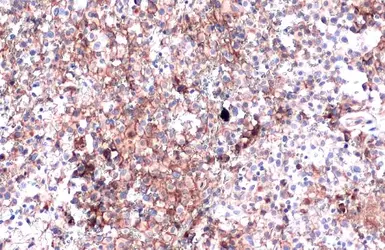
PD-L1 antibody detects PD-L1 protein at cell membrane by immunohistochemical analysis.
Sample: Paraffin-embedded human ovarian cancer.
PD-L1 stained by PD-L1 antibody (GTX104763) diluted at 1:4000.
Antigen Retrieval: Citrate buffer, pH 6.0, 15 min

Anti-PD-L1 antibody used in Immunocytochemistry/ Immunofluorescence (ICC/IF). GTX104763

GTX104763 ICC/IF Image

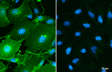
PD-L1 antibody detects PD-L1 protein by immunofluorescent analysis.
Sample: MDA-MB-231 (left) and HeLa (right) cells were fixed in ice-cold MeOH for 5 min.
Green: PD-L1 stained by PD-L1 antibody (GTX104763) diluted at 1:500.
Blue: Hoechst 33342 staining.
Scale bar= 10 μm.

Anti-PD-L1 antibody used in IHC (Paraffin sections) (IHC-P). GTX104763

GTX104763 IHC-P Image

PD-L1 antibody detects PD-L1 proteinat cell membrane in human ovarian carcinoma by immunohistochemical analysis.
Sample: Paraffin-embedded human ovarian carcinoma.
PD-L1 antibody (GTX104763) diluted at 1:1000.

Antigen Retrieval: Citrate buffer, pH 6.0, 15 min

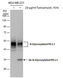
Anti-PD-L1 antibody used in Western Blot (WB). GTX104763

GTX104763 WB Image

Untreated (–) and treated (+) MDA-MB-231 whole cell extracts (30 μg) were separated by 10% SDS-PAGE, and the membrane was blotted with PD-L1 antibody (GTX104763) diluted at 1:1000.

Anti-PD-L1 antibody used in Western Blot (WB). GTX104763

GTX104763 WB Image

Various whole cell extracts (30 μg) were separated by 12% SDS-PAGE, and the membranes were blotted with PD-L1 antibody (GTX104763) diluted at 1:2000 and competitor's antibody (CST#13684) diluted by 1:500. The HRP-conjugated anti-rabbit IgG antibody (GTX213110-01) was used to detect the primary antibody.
*The competitor is not affiliated with GeneTex and does not endorse this product.
Anti-PD-L1 antibody used in IHC (Paraffin sections) (IHC-P). GTX104763

GTX104763 IHC-P Image

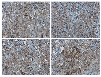
PD-L1 antibody detects PD-L1 protein at cell membrane in PD-L1 protein-expressing cell lines by immunohistochemical analysis. Antibodies: PD-L1 antibody (GTX104763) diluted at 1:1000, and competitor's antibody diluted at 1:50. Samples: Negative (-), low positive (+), intermediate positive (++) and strong positive (+++) cell line cores assessed using Quantitative Digital Pathology.
Antigen Retrieval: Citrate buffer, pH 6.0, 15 min
*The competitor is not affiliated with GeneTex and does not endorse this product.

Anti-PD-L1 antibody used in IHC (Paraffin sections) (IHC-P). GTX104763

GTX104763 IHC-P Image

PD-L1 antibody detects PD-L1 protein at cell membrane in human ovarian carcinoma by immunohistochemical analysis. Antibodies: PD-L1 antibody (GTX104763) diluted at 1:1000, and competitor's antibody diluted at 1:50.
Antigen Retrieval: Citrate buffer, pH 6.0, 15 min
*The competitor is not affiliated with GeneTex and does not endorse this product.

Anti-PD-L1 antibody used in Western Blot (WB). GTX104763

GTX104763 WB Image

Non-transfected (–) and transfected (+) 293T whole cell extracts (30 μg) were separated by 10% SDS-PAGE, and the membrane was blotted with PD-L1 antibody (GTX104763) diluted at 1:1000. The HRP-conjugated anti-rabbit IgG antibody (GTX213110-01) was used to detect the primary antibody, and the signal was developed with Trident ECL plus-Enhanced.

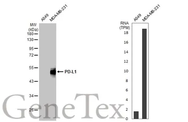
Anti-PD-L1 antibody used in Western Blot (WB). GTX104763

GTX104763 WB Image

Various whole cell extracts (30 μg) were separated by 10% SDS-PAGE, and the membrane was blotted with PD-L1 antibody (GTX104763) diluted at 1:2000. The HRP-conjugated anti-rabbit IgG antibody (GTX213110-01) was used to detect the primary antibody. Corresponding RNA expression data for the same cell lines are based on Human Protein Atlas program.
REFERENCE

REVIEW

PD-L1 antibody Cat. No. GTX104763

Rating ( Average 4.8 based on 11 users reviews)
Application
Western Blot(WB) ( Average 4.8 based on 11 users reviews)
Date : Anonymous submitted on 24-Jul-2023
Score :
Application Tested : WB
Sample Species : Hu
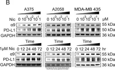
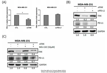
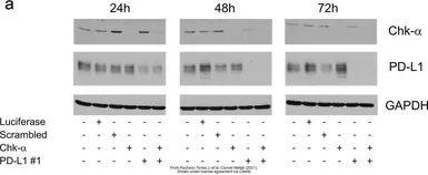
Figure Note : The data was peer reviewed and published in the 2021 in Cancer Metab. PMID: 33608051
Date : Anonymous submitted on 10-Aug-2021
Score :
Application Tested : WB
Sample Species : Hu
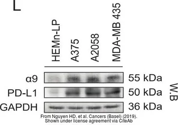
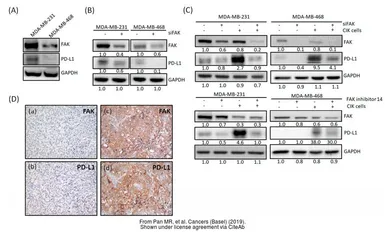
Figure Note : The data was peer reviewed and published in the journal Cancers (Basel) in 2019.PMID: 31835799
Date : Anonymous submitted on 10-Aug-2021
Score :
Application Tested : WB
Sample Species : Hu
Figure Note : The data was peer reviewed and published in the journal Cancers (Basel) in 2019.PMID: 31835799
Date : Anonymous submitted on 10-Aug-2021
Score :
Application Tested : WB
Sample Species : Hu
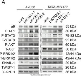
Figure Note : The data was peer reviewed and published in the journal Cancers (Basel) in 2019.PMID: 31835799
Date : Anonymous submitted on 10-Aug-2021
Score :
Application Tested : WB
Sample Species : Hu
Figure Note : The data was peer reviewed and published in the journal Cancers (Basel) in 2019.PMID: 31835799
Date : Anonymous submitted on 10-Aug-2021
Score :
Application Tested : WB
Sample Species : Hu
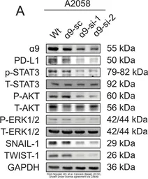
Figure Note : The data was peer reviewed and published in the journal Cancers (Basel) in 2019.PMID: 31905966
Date : Anonymous submitted on 10-Aug-2021
Score :
Application Tested : WB
Sample Species : Hu
Figure Note : The data was peer reviewed and published in the journal Cancers (Basel) in 2019.PMID: 31905966
Date : Anonymous submitted on 09-May-2021
Score :
Application Tested : WB
Sample Species : Hu
Sample : Huh7 cells
Amount used : 10 μg
Blocking : 5% skim milk in TBST, 27°C, 1Hr
Primary Antibody : 1:5000, 4°C, 12Hr
Secondary Antibody : GTX213110-01, 1:5000, 27°C, 2Hr
Lane Description : Two groups (regorafenib -/+) of whole cell extracts (10 μg) were separated by 12% SDS-PAGE, and the membrane was blotted with PD-L1 antibody (GTX104763) diluted at 1:5000. The HRP-conjugated anti-rabbit IgG antibody (GTX213110-01) was used to detect the primary antibody.
Date : Anonymous submitted on 18-Mar-2021
Score :
Application Tested : WB
Sample Species : Hu
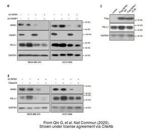
Figure Note : The data was peer reviewed and published in the journal Nat Commun in 2020. PMID: 32245950
Date : Anonymous submitted on 18-Mar-2021
Score :
Application Tested : WB
Sample Species : Hu
Figure Note : The data was peer reviewed and published in the journal Nat Commun in 2020. PMID: 32245950
Date : Anonymous submitted on 22-May-2017
Score :
Application Tested : WB
Sample Species : Hu
Sample : SK-LMS-1 cells
Amount used : 30 μg
Blocking : 3%BSA, 25°C, 0.5Hr
Primary Antibody : 1:1000, 4°C, 16Hr
Application Reference
SDS
PBS.pdf
Glycerol.pdf
Proclin.pdf
Package List Price ($)
$ 399
$ 169