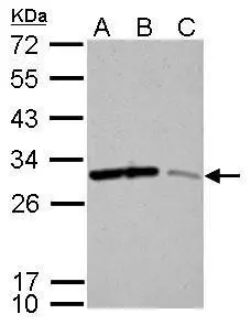
14-3-3 sigma antibody

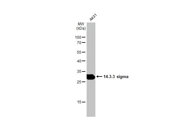
Whole cell extract (30 μg) was separated by 12% SDS-PAGE, and the membrane was blotted with 14-3-3 sigma antibody (GTX100801) diluted at 1:2000. The HRP-conjugated anti-rabbit IgG antibody (GTX213110-01) was used to detect the primary antibody.
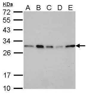
14-3-3 sigma antibody detects SFN protein by western blot analysis.
A. 30 μg 293T whole cell lysate/extract
B. 30 μg A431 whole cell lysate/extract
C. 30 μg HeLa whole cell lysate/extract
D. 30 μg HepG2 whole cell lysate/extract
E. 30 μg A375 whole cell lysate/extract
12% SDS-PAGE
14-3-3 sigma antibody (GTX100801) dilution: 1:2000
The HRP-conjugated anti-rabbit IgG antibody (GTX213110-01) was used to detect the primary antibody.
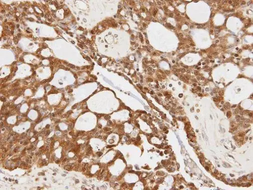
Immunohistochemical analysis of paraffin-embedded OVCA xenograft, using 14-3-3 sigma(GTX100801) antibody at 1:100 dilution.
Antigen Retrieval: Trilogy™ (EDTA based, pH 8.0) buffer, 15min
Whole cell extract (30 μg) was separated by 12% SDS-PAGE, and the membrane was blotted with 14-3-3 sigma antibody (GTX100801) diluted at 1:2000. The HRP-conjugated anti-rabbit IgG antibody (GTX213110-01) was used to detect the primary antibody.
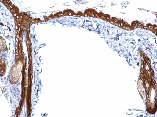
14-3-3 sigma antibody detects 14-3-3 sigma protein at cytosol on mouse skin by immunohistochemical analysis.
Sample: Paraffin-embedded mouse skin.
14-3-3 sigma antibody (GTX100801) dilution: 1:500.
Antigen Retrieval: Trilogy™ (EDTA based, pH 8.0) buffer, 15min
14-3-3 sigma antibody immunoprecipitates SFN protein in IP experiments.
IP samples: A431 whole cell extract
A. 40 μg A431 whole cell extract
B. Control with 4 μg of preimmune Rabbit IgG
C. Immunoprecipitation of SFN protein by 4 μg 14-3-3 sigma antibody (GTX100801)
5 % SDS-PAGE
The immunoprecipitated SFN protein was detected by 14-3-3 sigma antibody (GTX100801) diluted at 1:500.
[EasyBlot anti-rabbit IgG (GTX221666-01) was used as a secondary reagent]
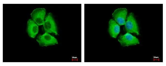
14-3-3 sigma antibody detects 14-3-3 sigma protein at cytoplasm by immunofluorescent analysis.
Sample: A431 cells were fixed in 4% paraformaldehyde at RT for 15 min.
Green: 14-3-3 sigma protein stained by 14-3-3 sigma antibody (GTX100801) diluted at 1:500.
Blue: Hoechst 33343 staining.
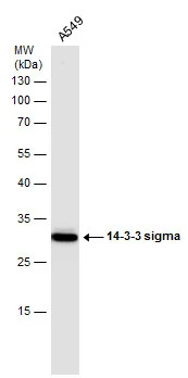
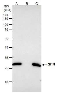
14-3-3 sigma antibody detects SFN protein by western blot analysis.
A. 30 μg Neuro2A whole cell lysate/extract
B. 30 μg GL261 whole cell lysate/extract
C. 30 μg C8D30 whole cell lysate/extract
12% SDS-PAGE
14-3-3 sigma antibody (GTX100801) dilution: 1:5000
The HRP-conjugated anti-rabbit IgG antibody (GTX213110-01) was used to detect the primary antibody.
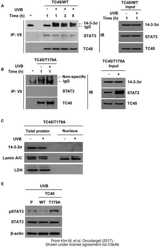
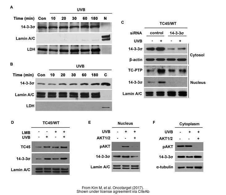
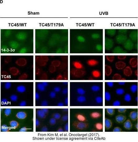
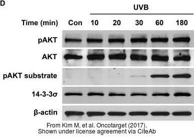
The data was published in the journal Oncotarget in 2017. PMID: 29207596
-
HostRabbit
-
ClonalityPolyclonal
-
IsotypeIgG
-
ApplicationsWB ICC/IF IHC-P IP
-
ReactivityHuman, Mouse