ABAT antibody

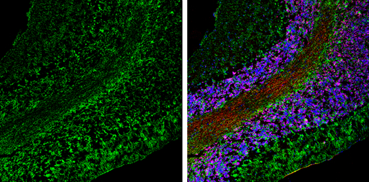
ABAT antibody detects ABAT Protein expression by immunohistochemical analysis.
Sample: Frozen-sectioned adult mouse cerebellum.
Green: ABAT stained by ABAT antibody (GTX133362) diluted at 1:250.
Red: NeuN, stained by NeuN antibody [2Q158] (GTX30773) diluted at 1:500.
Blue: Fluoroshield with DAPI (GTX30920).
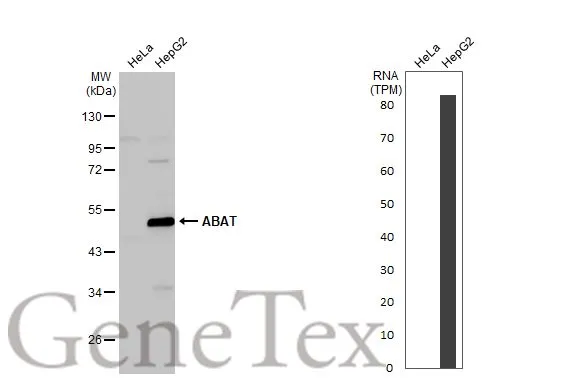
Various whole cell extracts (30 μg) were separated by 10% SDS-PAGE, and the membrane was blotted with ABAT antibody (GTX133362) diluted at 1:1000. The HRP-conjugated anti-rabbit IgG antibody (GTX213110-01) was used to detect the primary antibody. Corresponding RNA expression data for the same cell lines are based on Human Protein Atlas program.
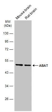
Various tissue extracts (10 μg) were separated by 10% SDS-PAGE, and the membrane was blotted with ABAT antibody (GTX133362) diluted at 1:5000.
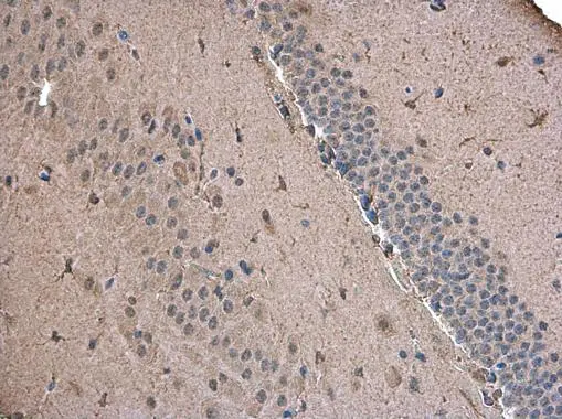
ABAT antibody detects ABAT protein at cytoplasm in mouse brain by immunohistochemical analysis.
Sample: Paraffin-embedded mouse brain.
ABAT antibody (GTX133362) diluted at 1:500.
-
HostRabbit
-
ClonalityPolyclonal
-
IsotypeIgG
-
ApplicationsWB IHC-P IHC-Fr
-
ReactivityHuman, Mouse, Rat