ACSL3 antibody
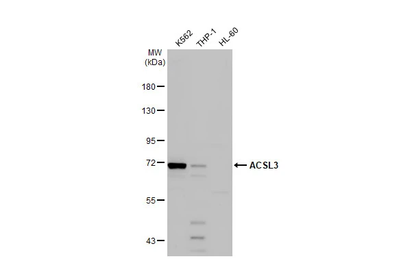
Various whole cell extracts (30 μg) were separated by 7.5% SDS-PAGE, and the membrane was blotted with ACSL3 antibody (GTX112431) diluted at 1:1000. The HRP-conjugated anti-rabbit IgG antibody (GTX213110-01) was used to detect the primary antibody.
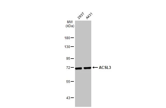
Various whole cell extracts (30 μg) were separated by 7.5% SDS-PAGE, and the membrane was blotted with ACSL3 antibody (GTX112431) diluted at 1:1000. The HRP-conjugated anti-rabbit IgG antibody (GTX213110-01) was used to detect the primary antibody.
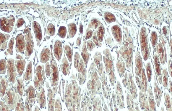
ACSL3 antibody detects ACSL3 protein at mitochondria by immunohistochemical analysis.
Sample: Paraffin-embedded mouse stomach.
ACSL3 stained by ACSL3 antibody (GTX112431) diluted at 1:500.
Antigen Retrieval: Citrate buffer, pH 6.0, 15 min
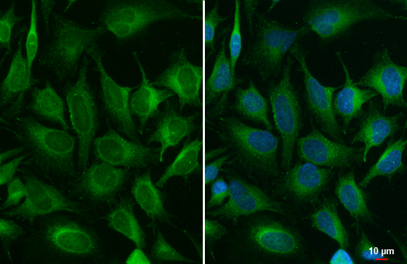
ACSL3 antibody detects ACSL3 protein at endoplasmic reticulum by immunofluorescent analysis.Sample: HeLa cells were fixed in 4% paraformaldehyde at RT for 15 min.Green: ACSL3 stained by ACSL3 antibody (GTX112431) diluted at 1:500.
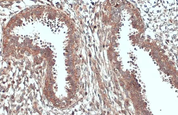
Immunohistochemical analysis of paraffin-embedded human breast cancer, using ACSL3(GTX112431) antibody at 1:500 dilution.
Antigen Retrieval: Trilogy™ (EDTA based, pH 8.0) buffer, 15min
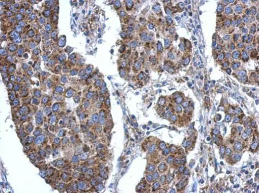
ACSL3 antibody detects ACSL3 protein at cytoplasm by immunohistochemical analysis.Sample: Paraffin-embedded human breast carcinoma.ACSL3 stained by ACSL3 antibody (GTX112431) diluted at 1:500.Antigen Retrieval: Citrate buffer, pH 6.0, 15 min
ACSL3 antibody detects ACSL3 protein at mitochondria by immunohistochemical analysis.
Sample: Paraffin-embedded mouse stomach.
ACSL3 stained by ACSL3 antibody (GTX112431) diluted at 1:500.
Antigen Retrieval: Citrate buffer, pH 6.0, 15 min
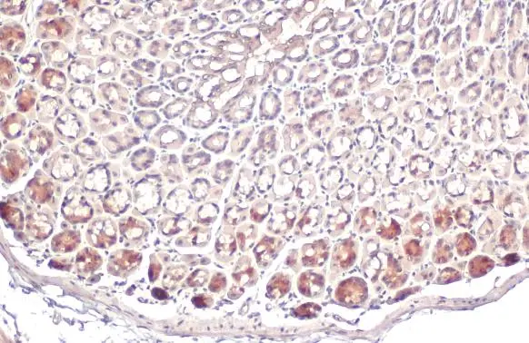
ACSL3 antibody detects ACSL3 protein at cytoplasm by immunohistochemical analysis.
Sample: Paraffin-embedded mouse stomach.
ACSL3 stained by ACSL3 antibody (GTX112431) diluted at 1:200.
Antigen Retrieval: Citrate buffer, pH 6.0, 15 min

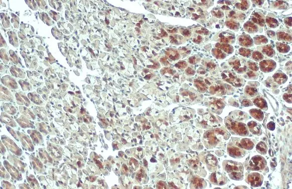
ACSL3 antibody detects ACSL3 protein at cytoplasm by immunohistochemical analysis.
Sample: Paraffin-embedded rat colon.
ACSL3 stained by ACSL3 antibody (GTX112431) diluted at 1:200.
Antigen Retrieval: Citrate buffer, pH 6.0, 15 min
-
HostRabbit
-
ClonalityPolyclonal
-
IsotypeIgG
-
ApplicationsWB ICC/IF IHC-P
-
ReactivityHuman, Mouse, Rat